

High prevalence of pineal cysts in healthy adults demonstrated by high-resolution, noncontrast brain MR imaging
- PMID: 17885233
- PMCID: PMC8134222
- DOI: 10.3174/ajnr.A0656
High prevalence of pineal cysts in healthy adults demonstrated by high-resolution, noncontrast brain MR imaging
Abstract
Background and purpose: Although the prevalence of pineal cysts in autopsy series has been reported as being between 25% and 40%, MR studies have documented their frequency to range between 1.5% and 10.8%. The purpose of this high-resolution brain MR imaging study at 1.9T was to determine the prevalence of pineal cysts in healthy adults.
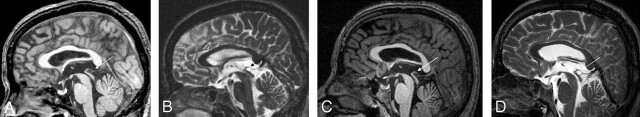
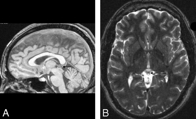
Materials and methods: Brain MR images of 100 healthy young volunteers were randomly selected from our International Consortium for Brain Mapping project data base. Cysts were detected as circular areas of isointensity relative to CSF on both 3D gradient-echo T1-weighted and 2D fast spin-echo T2-weighted images. The inner diameters of all visualized pineal cysts were measured, and a criterion of 2.0 mm of the largest inner cross-sectional diameter was used to categorize cysts as being either small cystic changes (<2.0-mm diameter) or pineal cysts (>2.0-mm diameter).
Results: Twenty-three percent (23/100) of the volunteers had pineal cysts with a mean largest inner cross-sectional diameter of 4.3 mm (range, 2-14 mm); 13% (13/100) demonstrated cystic changes involving the pineal gland with the largest inner cross-sectional diameter of less than 2.0 mm. There was a slight female predominance. Two subjects with long-term follow-up scans showed no symptoms or changes in the size of their pineal cysts.
Conclusion: On high-resolution MR imaging, the prevalence of pineal cysts was 23% in our healthy group of adults, which is consistent with previous autopsy studies. Long-term follow-up studies of 2 cases demonstrated the stability of the cysts.
Figures
References
-
- Tapp E, Huxley M. The histological appearance of the human pineal gland from puberty to old age. J Pathol 1972;108:137–44 - PubMed
-
- Tapp E. The histology and pathology of the human pineal gland. Prog Brain Res 1979;52:481–500 - PubMed
-
- Hasegawa A, Ohtsubo K, Mori W. Pineal gland in old age; quantitative and qualitative morphological study of 168 human autopsy cases. Brain Res 1987;409:343–49 - PubMed
-
- Hirato J, Nakazato Y. Pathology of pineal region tumors. J Neurooncol 2001;54:239–49 - PubMed
Publication types
MeSH terms
Grants and funding
LinkOut - more resources
Full Text Sources
Medical