

Improved identification of intracortical lesions in multiple sclerosis with phase-sensitive inversion recovery in combination with fast double inversion recovery MR imaging
- PMID: 17885241
- PMCID: PMC8134176
- DOI: 10.3174/ajnr.A0645
Improved identification of intracortical lesions in multiple sclerosis with phase-sensitive inversion recovery in combination with fast double inversion recovery MR imaging
Abstract
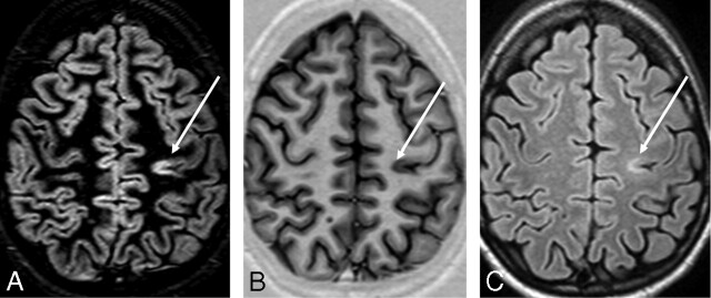
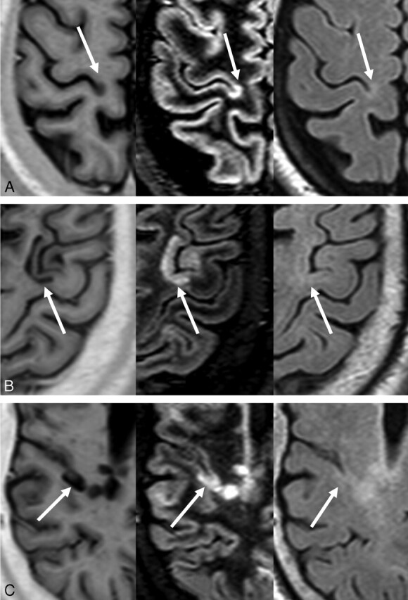
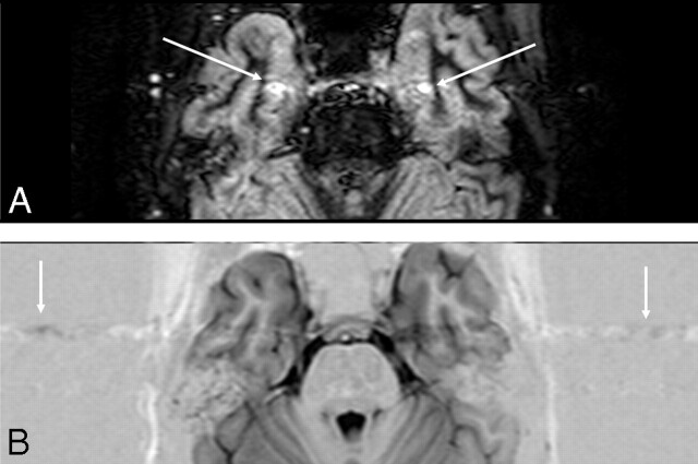
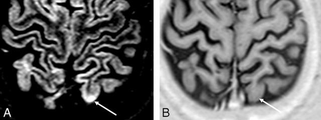
Background and purpose: Accurate detection and classification of purely intracortical lesions in multiple sclerosis (MS) are important in understanding their role in disease progression and impact on the clinical manifestations of the disease. However, detection of these lesions with conventional MR imaging remains a challenge. Although double inversion recovery (DIR) has been shown to improve the sensitivity of the detection of cortical lesions, this sequence has low signal-to-noise ratio (SNR), poor delineation of lesion borders, and is prone to image artifacts. We demonstrate that intracortical lesions can be identified and classified with greater confidence by the combination of DIR with phase-sensitive inversion recovery (PSIR) images.
Materials and methods: A total of 16 subjects with MS were included in this study. DIR, PSIR, and fluid-attenuated inversion recovery (FLAIR) images were acquired and inspected by 3 experts, with identification of lesions by consensus. PSIR and DIR images were jointly used to classify lesions as purely intracortical, mixed gray-white matter, and juxtacortical. The difference in the number of lesions detected in each category was compared between combined PSIR and DIR and conventional FLAIR.
Results: PSIR consistently allowed a clearer classification and delineation of lesions. Combined PSIR and DIR images showed a 337% improvement in the total number of lesions detected compared with FLAIR alone. Detection of intracortical lesions was improved by 417% compared with FLAIR. Detection of mixed gray-white matter and juxtacortical lesions was improved by 396% and 130%, respectively, compared with FLAIR.
Conclusion: Reliable detection and classification of intracortical lesions in MS are greatly improved by combined use of PSIR and DIR.
Figures
References
-
- Dawson JW. The histology of disseminated sclerosis. Trans Roy Soc Edinb 1916;50:517–740
-
- Lumsden CE. The neuropathology of multiple sclerosis. In: Vinken PJ, Bruyn GW, eds. Handbook of Clinical Neurology. Vol. 9. Amsterdam: Elsevier Science Publishers;1970. :217–309
-
- Bo L, Vedeler CA, Nyland HI, et al. Subpial demyelination in the cerebral cortex of multiple sclerosis patients. J Neuropathol Exp Neurol 2003;62:723–32 - PubMed
-
- Brink BP, Veerhuis R, Breij EC, et al. The pathology of multiple sclerosis is location-dependent: no significant complement activation is detected in purely cortical lesions. J Neuropathol Exp Neurol 2005;64:147–55 - PubMed
Publication types
MeSH terms
Grants and funding
LinkOut - more resources
Full Text Sources
Medical