. 2007 Oct;117(10):2952-61.
doi: 10.1172/JCI30639.

Ambient particulate matter accelerates coagulation via an IL-6-dependent pathway

Affiliations

Ambient particulate matter accelerates coagulation via an IL-6-dependent pathway

Gökhan M Mutlu et al. J Clin Invest. 2007 Oct.

Abstract

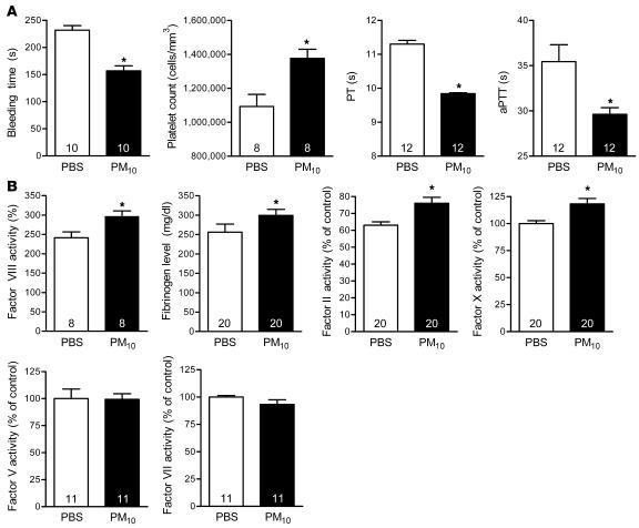
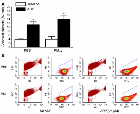
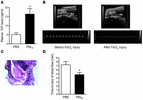
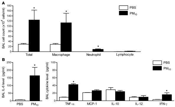
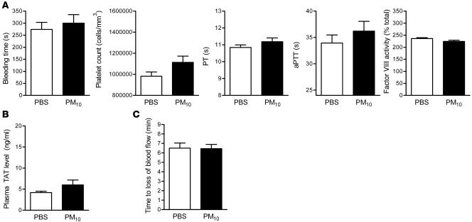
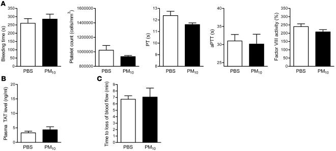
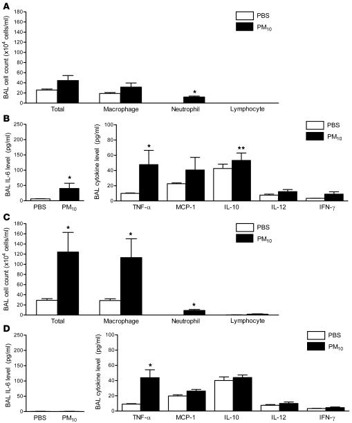
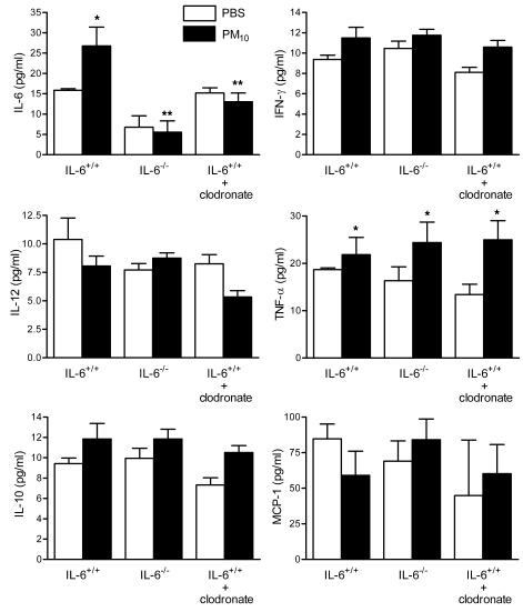
The mechanisms by which exposure to particulate matter increases the risk of cardiovascular events are not known. Recent human and animal data suggest that particulate matter may induce alterations in hemostatic factors. In this study we determined the mechanisms by which particulate matter might accelerate thrombosis. We found that mice treated with a dose of well characterized particulate matter of less than 10 microM in diameter exhibited a shortened bleeding time, decreased prothrombin and partial thromboplastin times (decreased plasma clotting times), increased levels of fibrinogen, and increased activity of factor II, VIII, and X. This prothrombotic tendency was associated with increased generation of intravascular thrombin, an acceleration of arterial thrombosis, and an increase in bronchoalveolar fluid concentration of the prothrombotic cytokine IL-6. Knockout mice lacking IL-6 were protected against particulate matter-induced intravascular thrombin formation and the acceleration of arterial thrombosis. Depletion of macrophages by the intratracheal administration of liposomal clodronate attenuated particulate matter-induced IL-6 production and the resultant prothrombotic tendency. Our findings suggest that exposure to particulate matter triggers IL-6 production by alveolar macrophages, resulting in reduced clotting times, intravascular thrombin formation, and accelerated arterial thrombosis. These results provide a potential mechanism linking ambient particulate matter exposure and thrombotic events.


Figures

Figure 1
Figure 1. Alterations in hemostasis in wild-type mice treated with PM.
Airborne PM10 (10 μg in 50 μl PBS) or PBS alone was intratracheally instilled into mouse lungs, and 24 hours later measurements of hemostasis were made. (A) Bleeding time, platelet count, PT, and aPTT measurements. (B) Fibrinogen and coagulation factors (factors II, V, VII, VIII, and X). The number inside each bar represents the number of animals for each group of experiments. *P < 0.05 for comparison between PM- and PBS-treated mice, n ≥ 8 in each treatment group.
Figure 2
Figure 2. Activation of platelets in mice treated with PM.
Airborne PM10 (10 μg in 50 μl PBS) or PBS alone was intratracheally instilled into mouse lungs, and 24 hours later platelet activation was measured via FACS analysis using CD62P antibody in the absence or presence of ADP. (A and B) Percent activation of platelets from mice exposed to PM10 or PBS (A) in the absence or presence of ADP (B). *P < 0.05 for comparison between PM- and PBS-treated mice, n ≥ 5 for each treatment group. fsc, forward scatter; ssc, side scatter.
Figure 3
Figure 3. Effect of PM on thrombin generation and arterial thrombosis.
(A) Plasma levels of TAT complexes in PM- and PBS-treated mice. (BD) Time to occlusion of the carotid artery after the application of FeCl3. (B) Representative 2D ultrasound view of the common carotid artery (CCA) and aorta (Ao) and Doppler from CCA before and after application of FeCl3 (at the time of cessation of blood flow). Loss of blood flow detected via Doppler was also associated with dilation of CCA. (C) Representative histology of CCA after FeCl3 injury showing a large thrombus with almost complete occlusion of arterial lumen (original magnification, ×200). (D) Time to loss of carotid blood flow in PM- or PBS-treated mice. *P < 0.05 for comparison between PM- and PBS-treated mice, n ≥ 4 in each treatment group.
Figure 4
Figure 4. Alterations in BAL fluid total cell count and differential and the levels of inflammatory cytokines in wild-type mice treated with PM.
(A) The total cell count and differential and (B) the levels of IL-6 and other inflammatory cytokines in the BAL fluid were measured 24 hours after the intratracheal instillation of PM10 or PBS. *P < 0.05 for comparison between PM- and PBS-treated mice, n ≥ 5 in each treatment group.
Figure 5
Figure 5. Effect of alveolar macrophage depletion on PM-induced alterations in hemostatic indices, thrombin generation, and arterial thrombosis.
(A) Measurements of hemostasis, including bleeding time, platelet count, PT, aPTT, and factor VIII activity. (B) TAT complex plasma levels. (C) Time to occlusion of the carotid artery after the application of FeCl3 in PM- and PBS-treated wild-type mice depleted of alveolar macrophages using liposomal clodronate. n ≥ 5 in each treatment group.
Figure 6
Figure 6. Effect of PM exposure on hemostatic indices, thrombin generation, and arterial thrombosis in IL6–/– mice.
(A) Measurements of hemostasis, including bleeding time, platelet count, PT, aPTT, and factor VIII activity. (B) TAT complex plasma levels. (C) Time to occlusion of the carotid artery after the application of FeCl3 in PM- and PBS-treated IL6–/– mice. n ≥ 5 in each treatment group.
Figure 7
Figure 7. Effect of PM exposure on BAL fluid total cell count and differential and the levels of inflammatory cytokines in IL6–/– mice and wild-type mice depleted of alveolar macrophages.
The total cell count and differential (A) and the levels of IL-6 and other inflammatory cytokines (B) in the BAL fluid were measured 24 hours after the intratracheal instillation of PM10 or PBS in wild-type mice treated with liposomal clodronate. (C and D) The same measurements were performed in IL6–/– mice. *P < 0.05 for comparison between PM and PBS treated mice; **P < 0.05 for comparison between sham-treated and liposomal clodronate–treated wild-type mice exposed to PM and between wild-type mice and IL6–/– mice exposed to PM. n ≥ 5 in each treatment group.
Figure 8
Figure 8. Effect of PM exposure on systemic cytokines.
Systemic levels of IL-6 and other inflammatory cytokines were measured 24 hours after the intratracheal instillation of PM10 or PBS in wild-type mice, IL6–/– mice, and wild-type mice with alveolar macrophage depletion (treated with liposomal clodronate 48 hours before treatment with PM or PBS). *P < 0.05 for comparison between PM- and PBS-treated mice, n ≥ 5 in each treatment group.

References

    1. Peters A., Dockery D.W., Muller J.E., Mittleman M.A. Increased particulate air pollution and the triggering of myocardial infarction. Circulation. 2001;103:2810–2815. - PubMed
    1. D’Ippoliti D., et al. Air pollution and myocardial infarction in Rome: a case-crossover analysis. Epidemiology. 2003;14:528–535. - PubMed
    1. Peters A., et al. Exposure to traffic and the onset of myocardial infarction. N. Engl. J. Med. 2004;351:1721–1730. - PubMed
    1. Rich D.Q., et al. Association of short-term ambient air pollution concentrations and ventricular arrhythmias. Am. J. Epidemiol. 2005;161:1123–1132. - PubMed
    1. Wellenius G.A., Bateson T.F., Mittleman M.A., Schwartz J. Particulate air pollution and the rate of hospitalization for congestive heart failure among Medicare beneficiaries in Pittsburgh, Pennsylvania. Am. J. Epidemiol. 2005;161:1030–1036. - PMC - PubMed

Publication types

MeSH terms