. 2007 Nov 1;311(1):25-39.
doi: 10.1016/j.ydbio.2007.08.003. Epub 2007 Aug 9.

Egfr/Ras signaling regulates DE-cadherin/Shotgun localization to control vein morphogenesis in the Drosophila wing

Affiliations

Egfr/Ras signaling regulates DE-cadherin/Shotgun localization to control vein morphogenesis in the Drosophila wing

David D O'Keefe et al. Dev Biol. .

Abstract

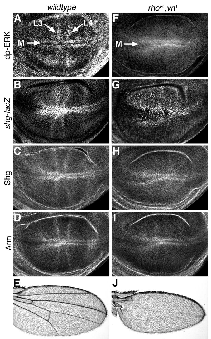
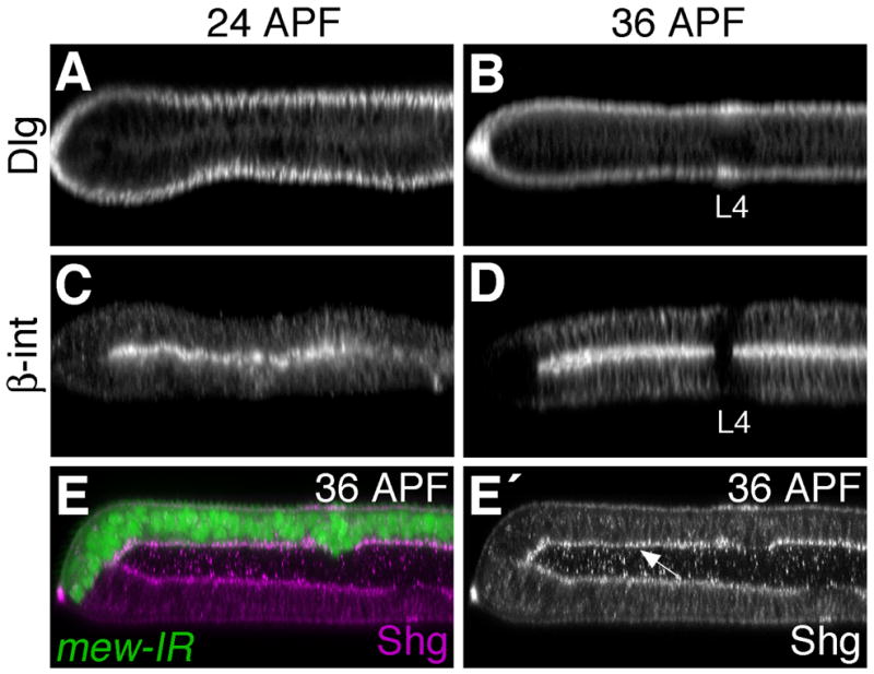
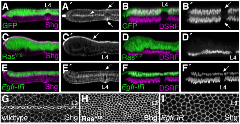
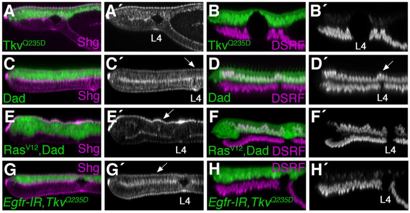
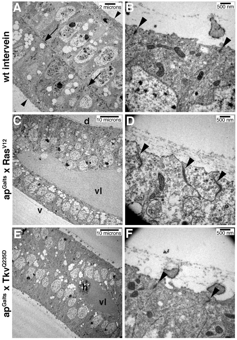
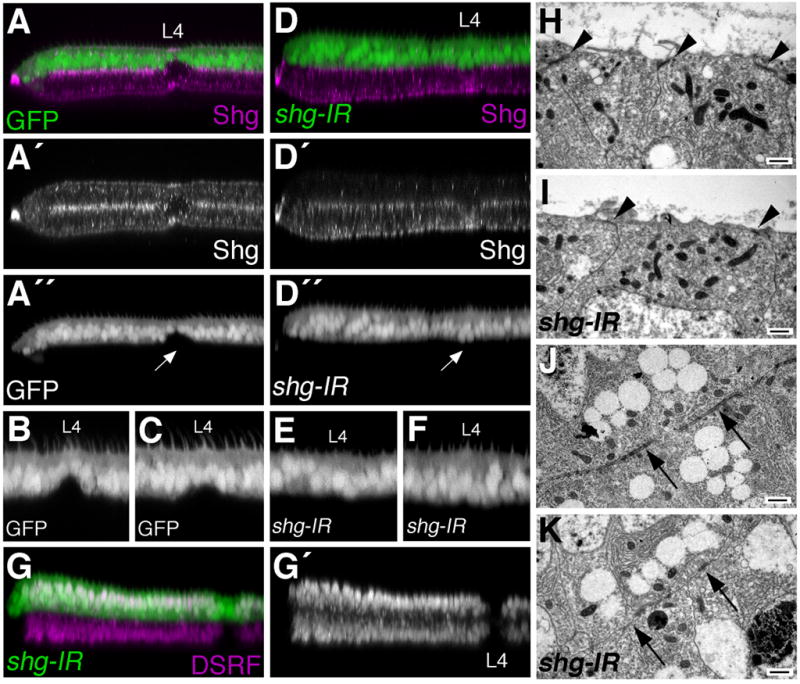
Egfr/Ras signaling promotes vein cell fate specification in the developing Drosophila wing. While the importance of Ras signaling in vein determination has been extensively documented, the mechanisms linking Ras activity to vein differentiation remain unclear. We found that Ras signaling regulates both the levels and subcellular localization of the cell adhesion molecule DE-cadherin/Shotgun (Shg) in the differentiating wing epithelium. High Ras activity in presumptive vein cells directs the apical localization of Shg containing adherens junctions, whereas low Ras activity in intervein cells allows Shg to relocalize basally. These alterations in Shg-mediated adhesion control cell shape changes that are essential for vein morphogenesis. While Decapentaplegic (Dpp) acts downstream of Ras to maintain vein cell identity in the pupal wing, our results indicate that Ras controls Shg localization via a Dpp-independent mechanism. Ras, therefore, regulates both the transcriptional responses necessary for vein cell identity, and the cell adhesive changes that determine vein and intervein cell morphology.


Figures

Fig. 1
Fig. 1. Ras signaling affects cell affinity and shg expression in the developing wing
Clones of cells expressing GFP are irregularly shaped (A). In contrast, clones of cells expressing RasV12 are rounded (B), suggesting they have altered adhesive properties and an inability to mix freely with neighboring wildtype cells. (C) The FLP/FRT technique was used to generate homozygous rasc40b mutant clones, marked by loss of GFP (arrows). Two copies of GFP mark wild-type twinspots, while heterozygous cells are marked by one copy of GFP. ras mutant clones survive poorly and are significantly rounded compared to their wildtype twinspots. (D) Large clones of cells lacking ras function were generated using the FLP/FRT, Minute technique (GFP-negative cells). These ras mutant cells have greatly reduced levels of shg expression, as measured by a shg-lacZ reporter construct. (E) Clones of cells expressing constitutively active RasV12 (GFP-positive cells) have higher levels of shg-lacZ expression.
Fig. 2
Fig. 2. Patterns of endogenous shg expression correlate with MAPK activity
(A) Ras/MAPK activity, as measured by di-phospho-ERK (dp-ERK) levels, is highest in presumptive vein and margin cells of the developing wing blade. Longitudinal veins L3 and L4, as well as the wing margin (M) are indicated. (B) shg expression is similarly highest in presumptive wing vein cells, as measured by a shg-lacZ reporter construct. Shg protein (C) and Arm protein (D) are also present in similar patterns. Late third instar wing discs from rhove,vn1 larvae have high MAPK activity in presumptive margin cells, but lack dp-ERK staining in presumptive veins (F). Similar patterns are present for shg-lacz (G), Shg protein (H), and Arm protein (I). Compared to wildtype adult wings (E), rhove,vn1 wings have a normal margin, but lack veins (J).
Fig. 3
Fig. 3. Shg localization is dynamically regulated during wing development
(A) In late third instar wildtype wing discs, Shg is found at highest levels in presumptive vein cells. Longitudinal veins L3 and L4, as well as the wing margin (M) are indicated. (A′) An optical cross section through the imaginal disc wing pouch indicates that the majority of Shg is near the apical cell surface (arrow). Arrowhead indicates the basal cell surface. (B–D) Pupal wings stained for Shg are shown. Proximal is to the left and anterior is up. Longitudinal vein L4 is indicated. (B′–C′) Optical cross sections through vein L4 and the posterior margin of pupal wings are shown. Posterior is to the left and vein L4 is indicated. At these stages of development the pupal wing is composed of two cell layers (dorsal and ventral) apposed at their basal cell surfaces. Arrows in B′ indicate the apical surfaces, while the arrowhead marks the basal surfaces. (B,B′) 24 hours after puparium formation (24 hours APF) Shg levels are highest in broad bands of presumptive vein cells, and the majority of Shg is sub-apically localized (B′). At 30 hours APF, Shg is found in more restricted bands of vein cells (C,C′), while Shg is also beginning to accumulate basally in intervein cells (C′). By 36 hours APF, sub-apical Shg is concentrated in narrow bands of vein cells (D, arrow D′), while high levels of Shg are found basally in intervein regions (arrowhead D′). Shg is also found at high levels in sensory organs of the margin and vein L3.
Fig. 4
Fig. 4. Apical/Basal polarity of intervein cells is maintained during early pupal stages
Discs large (A,B) and β-integrin (C,D), found apically and basally respectively, do not change their pattern of localization between 24 and 36 hours APF. (E) Loss of integrin function does not affect Shg localization. apGal4 was used to disrupt multiple edematous wings (mew) (the α-PS1-integrin subunit) function in dorsal wing cells (GFP-positive) using a UAS-mew-IR transgene. Wings were dissected at 36 hours APF and stained for Shg. While loss of mew function from early larval stages results in wing blistering (the dorsal and ventral wing surfaces are not apposed), the pattern of Shg localization is unaffected. Shg is still found near the basal surface of intervein cells (arrow E′).
Fig. 5
Fig. 5. Egfr/Ras signaling regulates Shg localization
(A–F,H,I) Using apGAL4 in combination with tubulin-GAL80ts, UAS-transgenes were activated in dorsal wing cells beginning at 0 hours APF. Wings were dissected at 36 hours APF and stained for either Shg (A,C,E,H,I) or the intervein marker DSRF (B,D,F). Optical cross-sections through the L4 vein region (A–F) or apical L3 (G–I) are shown. (A,B) In wings expressing GFP, dorsal and ventral cells are similar in appearance. Both dorsal and ventral vein cells (arrows B′) lack DSRF expression and have high levels of apical Shg (arrows A′). Intervein cells express DSRF and localize Shg to both apical and basal cell surfaces (arrowhead marks the basal surfaces). (C.D) RasV12 expression causes all dorsal wing cells to adopt the vein cell fate. DSRF expression is absent from RasV12, GFP positive cells (D). In these ectopic vein cells, Shg levels are elevated and the majority of the protein is apically localized (arrow C′). (E,F) Expression of an Egfr-IR transgene eliminates vein cells. DSRF localizes to all Egfr-IR expressing cells (arrow F′), and the apical accumulation of Shg in vein regions is not observed (arrow E′). (G–I) Apical surfaces of 36 hour APF pupal wings. Consistently magnified regions of the dorsal L3 wing vein are shown and Shg is visualized. (G) Wildtype vein and intervein cells are morphologically distinct. RasV12 expression (H) promotes apical constriction (as in vein cells), whereas Egfr-IR expression (I) promotes cell flattening (as in intervein cells).
Fig. 6
Fig. 6. Shg localization and vein cell fate are regulated independently
The apGalts system was used to express indicated transgenes from 0–36 hour APF. Wings were stained for Shg (A,C,E,G) or DSRF (B,D,F.H) and optical cross-sections through the L4 vein region are shown. An activated version of the Dpp receptor Thickvein (TkvQ235D) results in vein cell identity (B). While Shg is elevated in TkvQ235D expressing cells, the protein is not preferentially concentrated at the apical surface (A). Dad expression eliminates vein cell identity (D). Cells in the dorsal L4 vein region maintain DSRF expression (arrow D′) and adopt the intervein pattern of Shg localization (arrow C′). Expression of RasV12 together with Dad maintains the normal pattern of vein and intervein cell identity (F), but Shg levels are elevated and apically localized in RasV12, dad expressing intervein cells (arrow E′). In contrast, apical localization of Shg is not observed in Egfr-IR, TkvQ235D expressing vein cells (arrow G,H).
Fig. 7
Fig. 7. Ras signaling affects adherens junction morphology
The apGalts system was used to express GFP (A,B), RasV12 (C,D), or TkvQ235D (E,F) from 0–36 hours APF. Sections though 36 hour APF pupal wings imaged by TEM are shown. B,D,F are high magnification views of the apical, dorsal cell surface. (A) Arrows point to basal adhesion junctions between the dorsal and ventral wing epithelia. Arrowheads indicate the apical surfaces. (B) Arrowheads indicate adherens junctions between intervein cells. (C,D) RasV12 expression causes apico-basal extension of dorsal epithelial cells, wing blistering and enlarged apical adherens junctions (arrowheads D). (E,F) In contrast, TkvQ235D expression does not affect apical adherens junction morphology (arrowheads F). Vein lumen (vl), dorsal (d), ventral (v), haemocyte (h).
Fig. 8
Fig. 8. Loss of shg inhibits vein morphogenesis
apGal4 was used to express either GFP (A–C,G,I), or GFP together with a UAS-shg-IR transgene (D–F,H,J) in dorsal wing cells from 0–36 hours APF. (A–F) Optical cross-sections through vein L4 and the posterior margin of 36 hour APF wings are shown. Eliminating shg expression (compare A′ to D′) impairs the ability of vein cells to form a vein lumen (compare A,B,C to D,E,F). Arrow in A″ points to the L4 vein cells which have constricted along the apical/basal axis to form the dorsal portion of a vein lumen, while shg-ir expressing L4 vein cells do not contrict (arrow D″). Additional examples of dorsal L4 cells expressing GFP (B,C), or shg-IR (E,F) are shown. (G) Depletion of shg expression does not affect the pattern of vein/intervein identity, as measured by DSRF expression. (H–K) TEM analysis indicates that shg-IR expression affects adherens junction morphology. Apical adherens junctions found in control wings (arrowheads H) are nearly eliminated in shg-IR expressing cells (arrowheads I). Basal adherens junctions (arrows J) are similarly reduced (arrows K). Scale bars indicate 500nm.
Fig. 9
Fig. 9. Model of Egfr/Ras pathway function in early pupal vein cells
Rhomboid expression in presumptive vein cells activates Egfr ligands, leading to signaling through the Egfr/Ras pathway. Ras signaling directs vein cell identity through Dpp, and affects shg expression and protein localization (apical concentration) to alter vein cell shape in a Dpp-independent fashion.

References

    1. Andersson S, Saeboe-Larssen S, Lambertsson A, Merriam J, Jacobs-Lorena M. A Drosophila third chromosome Minute locus encodes a ribosomal protein. Genetics. 1994;137:513–20. - PMC - PubMed
    1. Baumgardt M, Miguel-Aliaga I, Karlsson D, Ekman H, Thor S. Specification of neuronal identities by feedforward combinatorial coding. PLoS Biol. 2007;5:e37. - PMC - PubMed
    1. Bier E. Localized activation of RTK/MAPK pathways during Drosophila development. Bioessays. 1998;20:189–94. - PubMed
    1. Brower DL, Jaffe SM. Requirement for integrins during Drosophila wing development. Nature. 1989;342:285–287. - PubMed
    1. Brown KE, Baonza A, Freeman M. Epithelial cell adhesion in the developing Drosophila retina is regulated by Atonal and the EGF receptor pathway. Dev Biol 2006 - PubMed

Publication types

LinkOut - more resources