Review
. 2007 Dec;29(4):303-9.
doi: 10.1016/j.jaut.2007.07.017. Epub 2007 Sep 20.

Innate and adaptive immune response to apoptotic cells

Affiliations
Review

Innate and adaptive immune response to apoptotic cells

Yufeng Peng et al. J Autoimmun. 2007 Dec.

Abstract

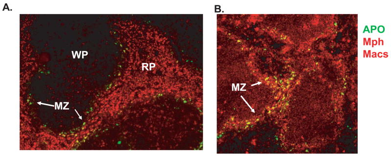
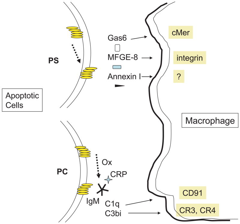
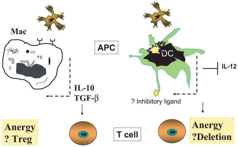
The immune system is constantly exposed to dying cells, most of which arise during central tolerance and from effete circulating immune cells. Under homeostatic conditions, phagocytes (predominantly macrophages and dendritic cells) belonging to the innate immune system, rapidly ingest cells and their debris. Apoptotic cell removal requires recognition of altered self on the apoptotic membrane, a process which is facilitated by natural antibodies and serum opsonins. Recognition, may be site and context specific. Uptake and ingestion of apoptotic cells promotes an immunosuppressive environment that avoids inflammatory responses to self-antigens. However, it does not preclude a T cell response and it is likely that constant exposure to self-antigen, particularly by immature dendritic cells, leads to T cell tolerance. Tolerance occurs by several different mechanisms including anergy and deletion (for CD8+T cells) and induction of T regulatory cells (for CD4+T cells). Failed apoptotic cell clearance promotes immune responses to self-antigens, especially when the cellular contents are leaked from the cell (necrosis). Inflammatory responses may be induced by nucleic acid stimulation of Toll like receptors and other immune sensors, specific intracellular proteins and non-protein (uric acid) stimulation of inflammasomes.


Figures

Figure 1
Figure 1. Apoptotic cells are removed from the circulation by macrophages in the marginal zone of the spleen
Apoptotic cells (labeled green) injected into normal mice are observed in the marginal zones in spleens harvested 4 hrs after injection. In A, cells were stained with anti-CD68 which recognizes all macrophage populations and in B, macrophages were stained with anti-CD169 which recognizes marginal metallophilic (Mph) macrophages. Frozen sections were analyzed by immunofluorescence at 10x magnification. WP= white pulp, RP =red pulp; MZ = marginal zone.
Figure 2
Figure 2. Role of modified lipids in the recognition and clearance of apoptotic cells
Upper left. Translocation of phosphatidylserine (PS) from the inside to outside of the apoptotic cell membrane allows serum opsonins to bind to PS. Different opsonins interact with different ligands on the phagocyte allowing compartment specific uptake to occur. Lower left. Phosphorylcholine, which is situated on the outside of the cell membrane may be biochemically modified in several ways on apoptotic cells. Caspase 3 activates the calcium independent phospholipase A2 (iPLA2) to cleave the fatty acid from PC, yielding lysoPLA2. LysoPLA2 acts as both an antigen for natural IgM antibodies as well as a chemoattractant for macrophages. PC may be oxidized by reactive oxygen (ROI) intermediates as well. Binding of natural antibodies to apoptotic cells explains, in part, activation of the classical pathway of complement and deposition of C3bi on the cell surface. C3bi serves as a ligand for CR3 and CR4 on phagocytes.
Figure 3
Figure 3. Effect of apoptotic cell ingestion on innate and adaptive immunity
Left panel. Ingestion of apoptotic cells by macrophages induces the expression of anti-inflammatory cytokines such as TGF-β and IL-10. Both of these cytokines are known to promote the expression of T regulatory cells (Treg). Right panel. Uptake of apoptotic cells by immature dendritic cells (iDC) leads to suppression of IL-12 production as well as possible changes in the cells surface expression of inhibitory ligands. Interaction of iDC with potentially autoreactive CD8+ T cells leads to anergy and/or deletion of CD8+ T cells. See text for details.

References

    1. Abbas AK, Lohr J, Knoechel B. Balancing autoaggressive and protective T cell responses. J Autoimmun. 2007;28:59–61. - PMC - PubMed
    1. Blank M, Shoenfeld Y. B cell targeted therapy in autoimmunity. J Autoimmun. 2007;28:62–8. - PubMed
    1. O’Brien BA, Geng X, Orteu CH, Huang Y, Ghoreishi M, Zhang Y, Bush JA, Li G, Finegood DT, Dutz JP. A deficiency in the in vivo clearance of apoptotic cells is a feature of the NOD mouse. J Autoimmun. 2006;26:104–15. - PubMed
    1. Greenberg ME, Sun M, Zhang R, Febbraio M, Silverstein R, Hazen SL. Oxidized phosphatidylserine-CD36 interactions play an essential role in macrophage-dependent phagocytosis of apoptotic cells. J Exp Med. 2006;203:2613–25. - PMC - PubMed
    1. Kim SJ, Gershov D, Ma X, Brot N, Elkon KB. I-PLA(2) activation during apoptosis promotes the exposure of membrane lysophosphatidylcholine leading to binding by natural immunoglobulin M antibodies and complement activation. J Exp Med. 2002;196:655–65. - PMC - PubMed

Publication types