

Mild chronic hypoxia modifies the fetal sheep neural and cardiovascular responses to repeated umbilical cord occlusion
- PMID: 17888891
- PMCID: PMC2078604
- DOI: 10.1016/j.brainres.2007.07.089
Mild chronic hypoxia modifies the fetal sheep neural and cardiovascular responses to repeated umbilical cord occlusion
Abstract
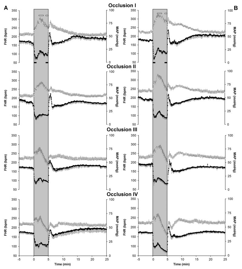
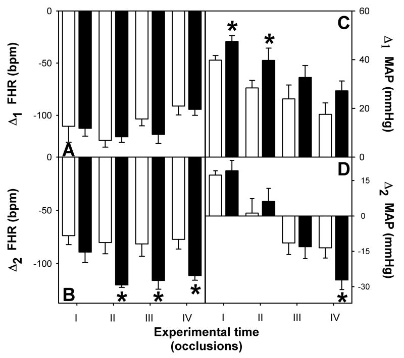
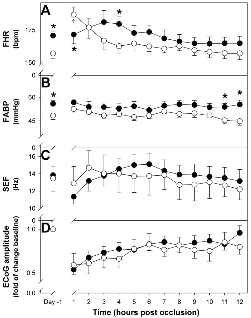
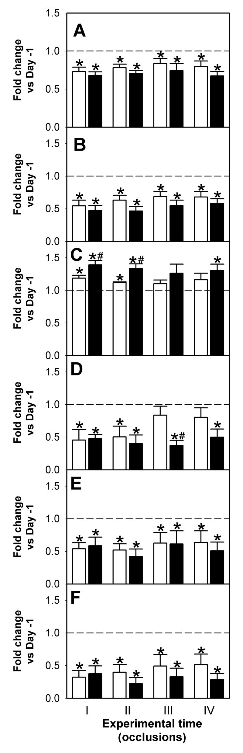
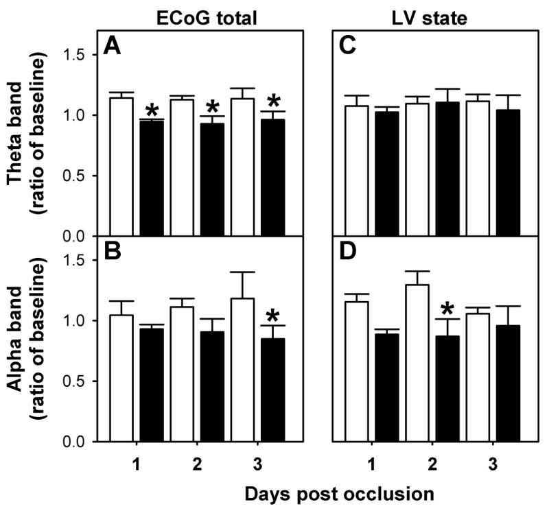
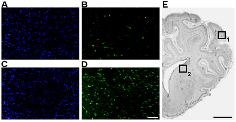
We have shown that 5 days of mild hypoxia has significant effects on fetal ECoG activity, heart rate and blood pressure. We now studied if mild prolonged hypoxemia had an adverse effect on the fetal cardiovascular and neural responses to repeated cord occlusion and on the magnitude of neuronal damage. Fetal and maternal catheters were placed at 120 days' gestation and animals allocated at random to receive intratracheal maternal administration of nitrogen (n=8) or compressed air in controls (n=7). Five days after surgery, nitrogen infusion was adjusted to reduce fetal brachial artery pO(2) by 25%. After 5 days of chronic hypoxemia, the umbilical cord was completely occluded for 5 min every 30 min for a total of four occlusions. Data are presented as mean+/-SEM and were analyzed by two-way ANOVA or two-sample t-test. Nitrogen infusion decreased fetal pO(2) by 26% (20.5+/-1.7 vs. 14.3+/-0.8 mm Hg) without changing fetal pCO(2) or pH. Pre-existing hypoxia fetuses had a greater terminal fall in heart rate in occlusions II, III and IV, and also had a more severe terminal hypotension in the final occlusion. Pre-existing hypoxia was associated with a greater fall in spectral edge frequency during occlusions from 14.4+/-0.9 Hz to 6.9+/-0.4 Hz vs. 13.6+/-1.64 Hz to 10.6+/-0.77 Hz in controls, p<0.05. In addition, during the three-day post-occlusion period, the contribution of theta and alpha band frequencies to total ECoG activity was significantly lower in the pre-existing hypoxia fetuses (p<0.05). These effects were associated with increased neuronal loss in the striatum (p<0.05). In summary, the cardiovascular and neural response indicates a detrimental effect of pre-existing mild hypoxia on fetal outcome following repeated umbilical cord occlusions.
Figures
Similar articles
-
Mild chronic hypoxemia modifies expression of brain stem angiotensin peptide receptors and reflex responses in fetal sheep.Am J Physiol Regul Integr Comp Physiol. 2009 Aug;297(2):R446-52. doi: 10.1152/ajpregu.00023.2009. Epub 2009 Jun 10. Am J Physiol Regul Integr Comp Physiol. 2009. PMID: 19515988 Free PMC article.
-
Prolonged mild hypoxia alters fetal sheep electrocorticogram activity.J Soc Gynecol Investig. 2006 Sep;13(6):404-11. doi: 10.1016/j.jsgi.2006.05.007. Epub 2006 Jul 31. J Soc Gynecol Investig. 2006. PMID: 16879989
-
Effect of repetitive umbilical cord occlusions on neuronal brain activity measured by the cerebral function analyzing monitor and histologic outcome in immature fetal sheep.J Soc Gynecol Investig. 2000 Jul-Aug;7(4):218-23. J Soc Gynecol Investig. 2000. PMID: 10964020
-
Fetal acidosis and hypotension during repeated umbilical cord occlusions are associated with enhanced chemoreflex responses in near-term fetal sheep.J Appl Physiol (1985). 2005 Oct;99(4):1477-82. doi: 10.1152/japplphysiol.00431.2005. Epub 2005 Jun 23. J Appl Physiol (1985). 2005. PMID: 15976361
-
Preexisting hypoxia is associated with a delayed but more sustained rise in T/QRS ratio during prolonged umbilical cord occlusion in near-term fetal sheep.Am J Physiol Regul Integr Comp Physiol. 2007 Sep;293(3):R1287-93. doi: 10.1152/ajpregu.00373.2007. Epub 2007 Jul 25. Am J Physiol Regul Integr Comp Physiol. 2007. PMID: 17652358
Cited by
-
Association between exposure to air pollution during pregnancy and false positives in fetal heart rate monitoring.Sci Rep. 2017 Sep 29;7(1):12421. doi: 10.1038/s41598-017-12663-2. Sci Rep. 2017. PMID: 28963562 Free PMC article.
-
Fault and blame, insults to the perinatal brain may be remote from time of birth.Clin Perinatol. 2014 Mar;41(1):105-17. doi: 10.1016/j.clp.2013.10.006. Epub 2013 Dec 15. Clin Perinatol. 2014. PMID: 24524449 Free PMC article. Review.
-
Fetal brain response to worsening acidosis: an experimental study in a fetal sheep model of umbilical cord occlusions.Sci Rep. 2023 Dec 27;13(1):23050. doi: 10.1038/s41598-023-49495-2. Sci Rep. 2023. PMID: 38155199 Free PMC article.
-
Mild chronic hypoxemia modifies expression of brain stem angiotensin peptide receptors and reflex responses in fetal sheep.Am J Physiol Regul Integr Comp Physiol. 2009 Aug;297(2):R446-52. doi: 10.1152/ajpregu.00023.2009. Epub 2009 Jun 10. Am J Physiol Regul Integr Comp Physiol. 2009. PMID: 19515988 Free PMC article.
-
Fetal in vivo continuous cardiovascular function during chronic hypoxia.J Physiol. 2016 Mar 1;594(5):1247-64. doi: 10.1113/JP271091. J Physiol. 2016. PMID: 26926316 Free PMC article.
References
-
- Aviado DM, Guevara AD. The Bezold-Jarisch reflex. A historical perspective of cardiopulmonary reflexes. Ann N Y Acad Sci. 2001;940:48–58. - PubMed
-
- Bartelds B, van BF, Teitel DF, Rudolph AM. Carotid, not aortic, chemoreceptors mediate the fetal cardiovascular response to acute hypoxemia in lambs. Pediatr Res. 1993;34:51–55. - PubMed
-
- Boekkooi PF, Baan J, Jr, Teitel D, Rudolph AM. Chemoreceptor responsiveness in fetal sheep. Am J Physiol. 1992;263:H162–H167. - PubMed
-
- Cohn HE, Sacks EJ, Heymann MA, Rudolph AM. Cardiovascular responses to hypoxemia and acidemia in fetal lambs. Am J Obstet Gynecol. 1974;120:817–824. - PubMed
Publication types
MeSH terms
Substances
Grants and funding
LinkOut - more resources
Full Text Sources
