. 2007 Sep 21;130(6):1108-19.
doi: 10.1016/j.cell.2007.07.013.

An intracellular serpin regulates necrosis by inhibiting the induction and sequelae of lysosomal injury

Affiliations

An intracellular serpin regulates necrosis by inhibiting the induction and sequelae of lysosomal injury

Cliff J Luke et al. Cell. .

Abstract

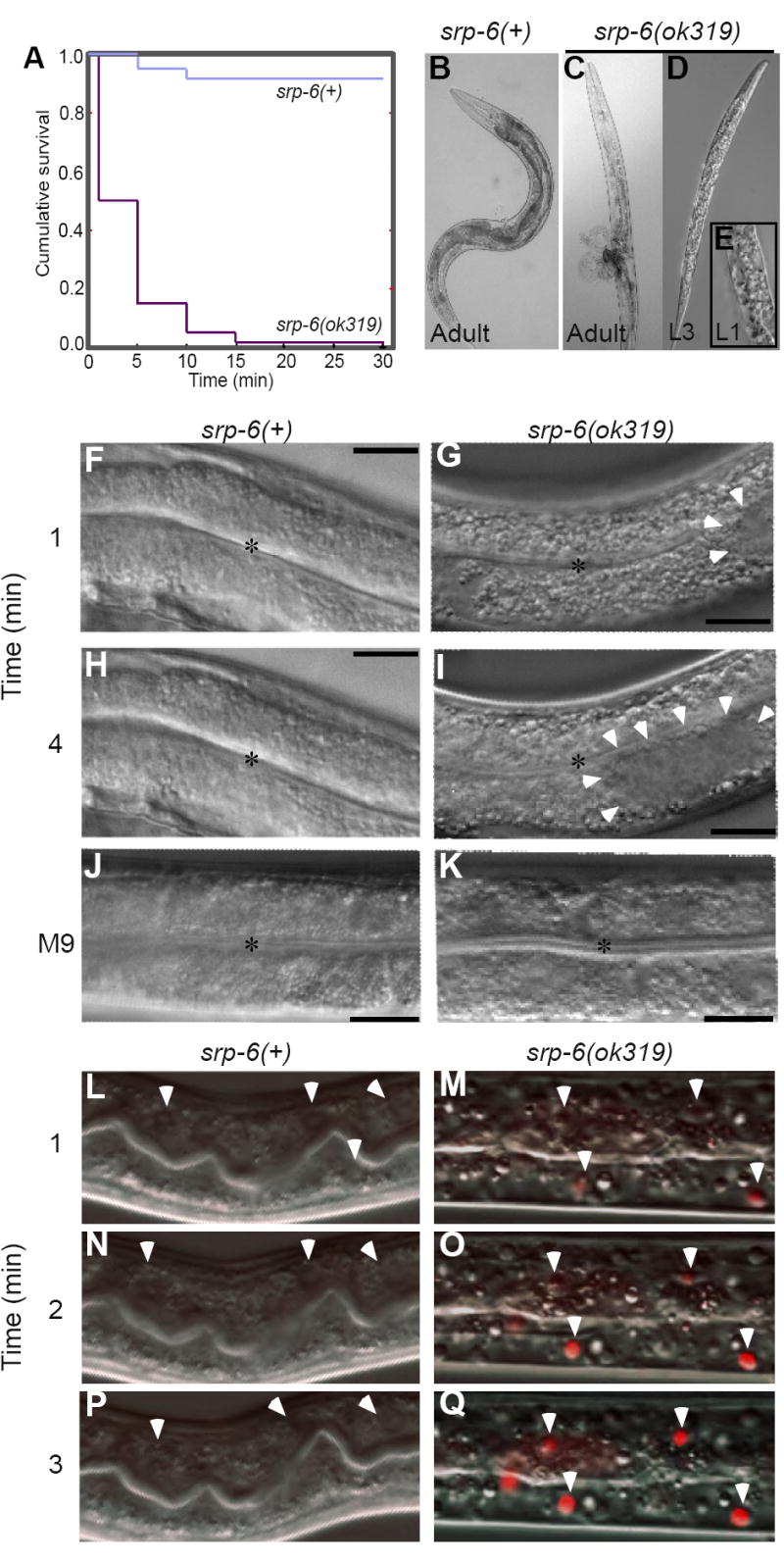
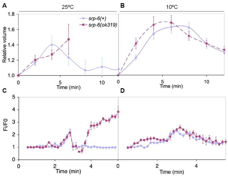
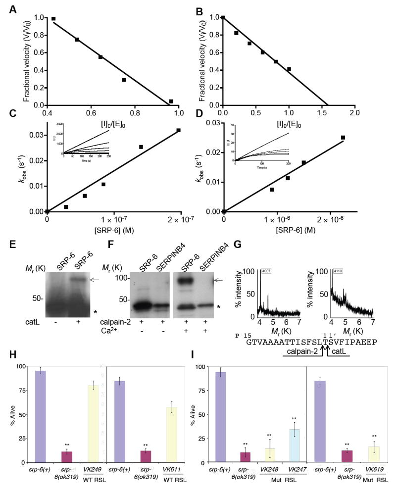
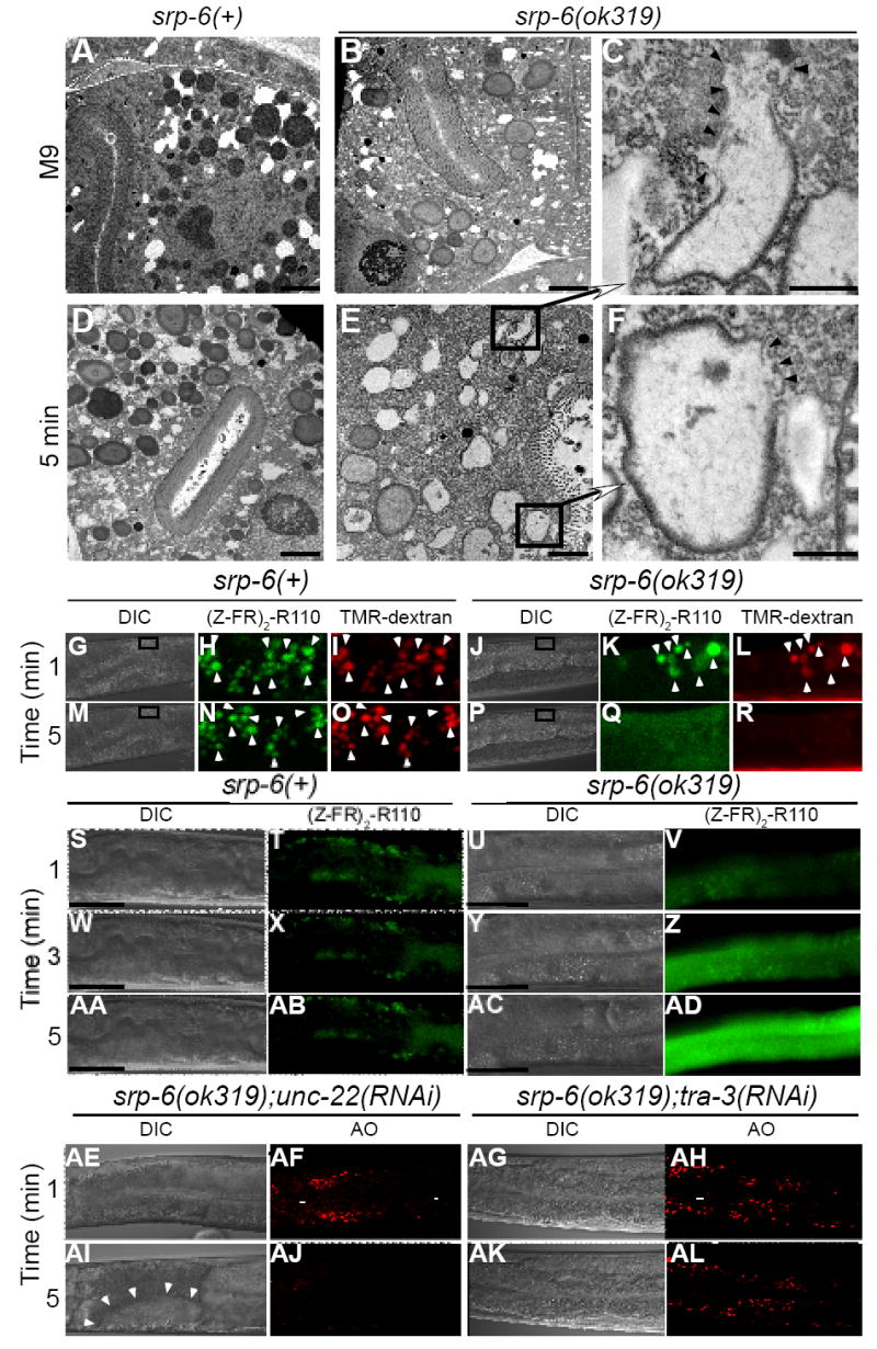
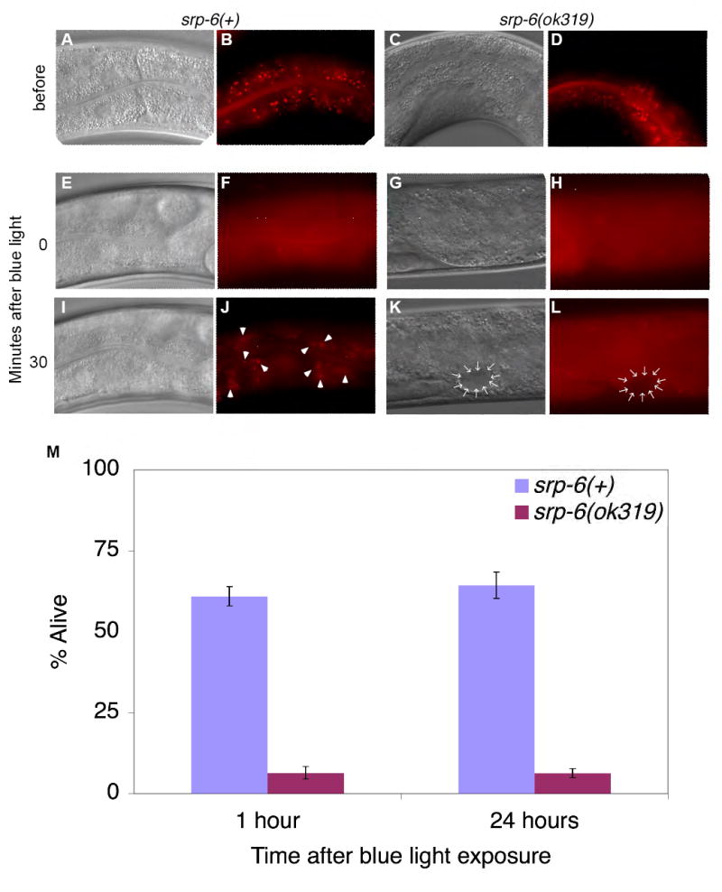
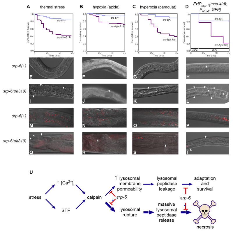
Extracellular serpins such as antithrombin and alpha1-antitrypsin are the quintessential regulators of proteolytic pathways. In contrast, the biological functions of the intracellular serpins remain obscure. We now report that the C. elegans intracellular serpin, SRP-6, exhibits a prosurvival function by blocking necrosis. Minutes after hypotonic shock, srp-6 null animals underwent a catastrophic series of events culminating in lysosomal disruption, cytoplasmic proteolysis, and death. This newly defined hypo-osmotic stress lethal (Osl) phenotype was dependent upon calpains and lysosomal cysteine peptidases, two in vitro targets of SRP-6. By protecting against both the induction of and the lethal effects from lysosomal injury, SRP-6 also blocked death induced by heat shock, oxidative stress, hypoxia, and cation channel hyperactivity. These findings suggest that multiple noxious stimuli converge upon a peptidase-driven, core stress response pathway that, in the absence of serpin regulation, triggers a lysosomal-dependent necrotic cell death routine.


Figures

Figure 1
Figure 1. srp-6 Nulls Displayed the Osl Phenotype
(A) Kaplan-Meier survival curves of srp-6(+) and srp-6(ok319) animals after immersion in 25 °C water (n ≈ 300 animals/group; P < 0.001, log-rank test). (B-K) Morphology of srp-6(+) (B, F, H, J) and srp-6(ok319) (C-E, G, I, K) animals during hypotonic stress. Low resolution DIC images of adults (B, C) and larvae (D, E) after 5 min in water. The L1 was fixed in 3% formaldehyde. For higher resolution DIC imaging (F-K; bar = 50 μm), L4s were immersed in water (F-I) or M9 (J, K). Intestinal cell cytoplasm of srp-6(ok319) animals was refractile (G, I) and clear cytoplasmic vacuoles (arrowheads) expanded and coalesced (*intestinal lumen). (L-Q) Intestinal cell plasma membrane integrity was lost in srp-6(ok319) (M, O, Q), but not in srp-6(+) (L, N, P) animals after exposure to water. Confocal microscopy was used to monitor propidium iodide uptake over time. Arrowheads indicate intestinal cell nuclei in dying animals.
Figure 2
Figure 2. srp-6(ok319) Animals Mounted a Regulatory Volume Decrease
(A-D) Relative changes in cell volume (A, B) and [Ca2+]i (C, D) over time in srp-6(+) (blue) and srp-6(ok319) (red) animals exposed to water at either 25 (A, C) or 10 °C (B, D). The means (± SD) were from a representative experiment (n = 8 animals/group).
Figure 3
Figure 3. Suppression of Osl
(A) Survival of ced-3(n717), ced-4(n1162) and ced-9(n1950) apoptosis mutants exposed to srp-6(RNAi) compared to control unc-22(RNAi). In all panels of this figure, mean survival (± SD) of young adults was assessed after exposure to water at 25 °C for 30 min. Asterisks indicate that the survival between groups was significantly different (n ≈ 300 animals/group, single and double asterisks indicate P < 0.05 and < 0.01, respectively). (B) Survival of srp-6(+) (blue) compared to srp-6(ok319) (red) animals pre-treated with caspase inhibitors, DEVD-CHO or YVAD-CHO, or the papain-like cysteine peptidase inhibitor, E-64d. (C) Survival of srp-6(+) (blue) compared to srp-6(ok319) (red) animals defective in autophagy by exposure to bec-1(RNAi) or empty vector(RNAi). Although bec-1(RNAi) did not suppress the Osl phenotype, this treatment did block normal dauer formation (not shown). (D) Survival of unc-68(kh30), itr-1(sa73) and crt-1(bz31) calcium-handling defective mutants exposed to srp-6(RNAi) compared to unc-22(RNAi). (E) Survival of srp-6(+) (blue) compared to srp-6(ok319) (red) animals defective in aspartic peptidase activity by exposure to asp-1(RNAi), asp-3(RNAi), asp-4(RNAi) or empty vector(RNAi). Note, survival of srp-6(ok319);empty vector (RNAi) compared to that of srp-6(ok319);asp-1(RNAi) was statistically different (P < 0.001). (F) Survival of srp-6(+) (blue) compared to srp-6(ok319) animals (red) defective in calpain peptidase activity by exposure to clp-1(RNAi), tra-3(RNAi), W05G11.4(RNAi) or empty vector(RNAi). Note, the tra-3(RNAi) and W05G11.4(RNAi) studies were conducted with those reported in (G), but are included in this panel to facilitate comparisons between the calpain(RNAi) studies. Thus, the same empty vector controls from (G) are also included in the panel reporting the tra-3(RNAi) findings. (G) Survival of srp-6(+) (blue) compared to srp-6(ok319) (red) animals defective in papain-like lysosomal cysteine peptidase activity by exposure to cysteine peptidase(RNAi) or empty vector(RNAi). (H) Survival of srp-6(+) or srp-6(ok319) animals defective in gut granule (lysosomal) biogenesis. The survival of srp-6(ok319), srp-6(+);glo-1(zu437) or srp-6(ok319);glo-1(zu437) animals was compared to that of srp-6(+) animals.
Figure 4
Figure 4. SRP-6 Inhibited Papain-like Lysosomal and Calpain Cysteine Peptidases
(A-B) The stoichiometry of inhibition (SI) for the interaction of SRP-6 with catL (A) or calpain-2 (B). Inhibitor:enzyme ratio of ~1:1 and ~1:1.5 completely inhibited catL and calpain-2 activity, respectively. SIs were determined from the mean of three separate experiments with representative plots shown. (C-D) The second-order rate constant (ka) for the interaction between SRP-6 and catL (C) or calpain-2 (D). The rate of inhibition (kobs) was measured over time (inset) and plotted against the molarity of the inhibitor to give a kapp of 9 × 10⁴ M-1 s-1 and 1.2 × 10⁴ M-1 s-1, respectively. Correcting for the enzymes Km for the substrate, yielded a ka of 10 × 104 ± 0.1 M-1 s-1 and 2.3 × 10⁴ ± 0.4 M-1 s-1 (mean ka ± SD from 3-4 experiments with representative plots shown), respectively. (E-F) Complex formation between mixtures of 35S-SRP-6 and catL (E) or calpain-2 (± 5 mM Ca2+) (F). Autoradiogram after SDS-PAGE revealed native or cleaved SRP-6 (*) and a higher molecular mass complex with catL or calpain-2 (arrow). SERPINB4 does not inhibit calpain-2. (G) The serpin reactive centers for the SRP-6-catL (left) and SRP-6-calpain-2 (right) interactions were deduced form inhibitor-enzyme complex cleavage fragments detected by MALDI-MS. The cleavage of SRP-6 occurred after the canonical P1 and P2 positions for catL and calpain-2, respectively (bottom). (H-I) Mean survival (± SD) in water of srp-6(+) or srp-6(ok319) animals rescued with a srp-6 extra-chromosomal array. Several transgenic lines were established with each construct, but only a representative line is shown. Animals were assayed for survival as described in Figure 3A (n ≈ 300 or ≈ 75 animals/group for transgenic lines under control of the srp-6 or nhx-2 promoter, respectively). (H) VK249 (srp-6(ok319);vkEx249[srp- 6(+);Pnhx-2∷GFP]) was established using a wild-type srp-6 gene and a co-injection marker, Pnhx-2∷GFP. VK611 (srp-6(ok319);vkEx611[Pnhx-2srp-6(+);Pnhx-2∷GFP]) was similar to VK249, except that srp-6 was driven by the intestinal cell specific promoter from nhx-2. Since the survival of srp-6(ok319) animals was unaffected by the Pnhx-2∷GFP transgene (not shown), only srp-6(ok319) survival data were reported. The survival of the srp-6(ok319) animals was compared to that of srp-6(+) animals. (I) VK248 (srp-6(ok319);vkEx248[srp-6(T327R);Pnhx-2∷GFP]), VK618 (srp-6(ok319);vkEx618[Pnhx-2srp-6(T327R);Pnhx-2∷GFP]) and VK247 (srp-6(ok319);vkEx247[srp-6(L339A;T340A)]) contained a extra-chromosomal array with either a P14 (T327R) or P2-P1 (L339A;T340A) RSL mutant srp-6.
Figure 5
Figure 5. Instability of Lysosomal Gut Granules in Osl
(A-F) Transmission electron microscopy of srp-6(+) (A, D) or srp-6(ok319) (B, E) animals 5 min after immersion in M9 (A, B) or water (D, E) (bar = 2 μm). Higher magnification (C, F) of lysosomal-like granules in (E) (bar = 500 nm). Arrowheads demarcate loss of granule membranes. (G-R) Live cell confocal microscopic imaging of the fluorescent cysteine peptidase substrate, (Z-FR)2-R110 (H, K, N, Q) and the fluorescent endocytic maker, TMR-dextran (I, L, O, R) in srp-6(+) (G-I, M-O) or srp-6(ok319) (J-L, P-R) animals after immersion in water. The fluorescent images were magnified from a DIC image (boxed inset) within the intestinal cell cytoplasm (G, M, J, P). The lysosomal-like gut granules of both srp-6(ok319) and srp-6(+) animals acquired both labels (arrowheads) and were mostly coincident in the merged images (Movies 5A and 5B). (S-AD) Live widefield DIC and fluorescence microscopy of srp-6(+) and srp-6(ok319) animals labelled with (Z-FR)2-R110 and immersed in water. Fluorescent gut lysosomes in the srp-6(+) animal remained intact (T, X, AB; bar = 50 μm). In the srp-6(ok319) animals, the disappearance of fluorescent lysosomes was accompanied by a transient wave of intense cytoplasmic fluorescence that propagated down the intestinal cell (V, Z, AD; Movies 6A and 6B). (AE-AL) Live DIC (AE, AG, AI, AK) and confocal microscopy (AF, AH, AJ, AL) of AO-labelled srp-6(ok319);unc-22(RNAi) (AE, AF, AI, AJ) or srp-6(ok319);tra-3(RNAi) (AG, AH, AK, AL) animals immersed in water. Vacuoles are indicated by arrowheads.
Figure 6
Figure 6. SRP-6 Protected Animals After Lysosomal Rupture
(A-L) Widefield DIC and fluorescence microscopy of a representative srp-6(+) (A, B) and srp-6(ok319) (C, D) animal labelled with the intestinal lysosomal marker, AO. After exposure to blue light, AO+ granules lysed in the srp-6(+) (F), and srp-6(ok319) (H) animals. The srp-6(ok319) animal also showed intestinal vacuolization (K, L; arrows). AO+ granules re-appeared (arrowheads) only in surviving srp-6(+)animals. (M) Mean (± SD; n ≈ 100 animals/group) survival of srp-6(+) and srp-6(ok319) animals treated with AO and blue light.
Figure 7
Figure 7. SRP-6 Protected Animals from Different Stressors
(A-D) Kaplan-Meier curves comparing the survival of srp-6(+) and srp-6(ok319) animals after exposure to (A) thermal stress, (B) hypoxia, (C) hyperoxia or (D) a Phsp-16mec-4(d) transgene. (n ≈ 75 animals/group; P < 0.001, log-rank test). (E-T) DIC (E-L) and fluorescent (M-T) images of intestinal cell vacuolization (arrowheads) and AO+ intestinal cell lysosomes (arrowheads), respectively, of srp-6(+) (E-H and M-P) and srp-6(ok319) animals (I-L and Q-T) exposed the stressor indicated above the survival curve. (E) Hypothetical core stress response pathway regulated by SRP-6. Different stressors converge on a core stress response pathway that triggers an increase in [Ca2+]i and modulation of at least one other stress-transducing factor (STF). Cytosolic Ca2+ and STF activate calpains, which associate with lysosomal membranes and enhance the lysosomal response to stress by facilitating, for example, autophagy. Calpains also increase lysosomal membrane permeability allowing for the small leakage of cysteine peptidases into the surrounding cytosol. Cytosolic cysteine peptidases also could provide an adaptive function by enhancing, for example, cytoskeletal rearrangements. However, in the absence of SRP-6, excessive calpain activity leads to massive lysosomal rupture, overwhelming release of unregulated lysosomal cysteine peptidases and necrotic cell death.

References

    1. Berger AJ, Hart AC, Kaplan JM. G alphas-induced neurodegeneration in Caenorhabditis elegans. J Neurosci. 1998;18:2871–2880. - PMC - PubMed
    1. Bolger BS, Dabbas M, Lopes A, Monaghan JM. Prognostic value of preoperative squamous cell carcinoma antigen level in patients surgically treated for cervical carcinoma. Gynecol Oncol. 1997;65:309–313. - PubMed
    1. Brenner S. The genetics of Caenorhabditis elegans. Genetics. 1974;77:71–94. - PMC - PubMed
    1. Chalfie M, Wolinsky E. The identification and suppression of inherited neurodegeneration in Caenorhabditis elegans. Nature. 1990;345:410–416. - PubMed
    1. Church FC, Pike RN, Tollefsen DM, Buckle AM, Ciaccia AV, Olson ST. Regulation of Hemostasis by Heparin-binding Serpins. In: Silverman GA, Lomas DA, editors. Molecular and Cellular Aspects of the Serpinopathies and Disorders of Serpin Activity. Singapore: World Scientific Publishing Co. Pte. Ltd.; 2007. pp. 509–554.

Publication types

MeSH terms