. 2007 Oct 22;426(3):145-8.
doi: 10.1016/j.neulet.2007.08.047. Epub 2007 Aug 25.

Oxygen glucose deprivation inhibits the growth and ERK phosphorylation of neural progenitor cells in vitro

Affiliations

Oxygen glucose deprivation inhibits the growth and ERK phosphorylation of neural progenitor cells in vitro

Haviryaji S G Kalluri et al. Neurosci Lett. .

Abstract

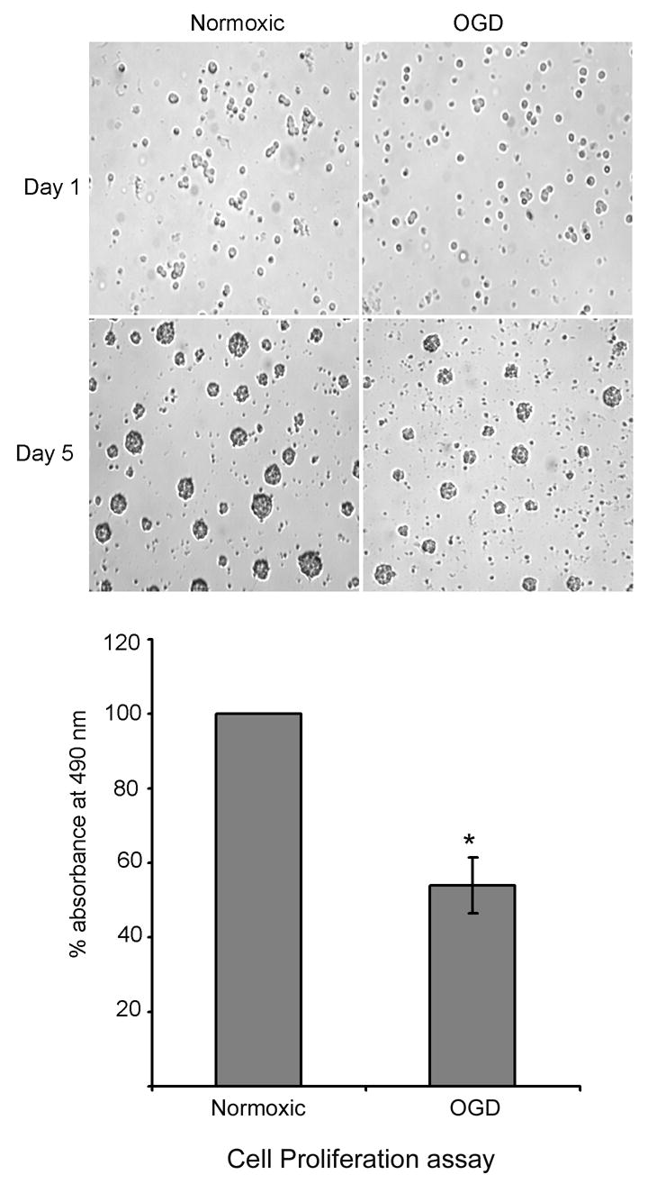
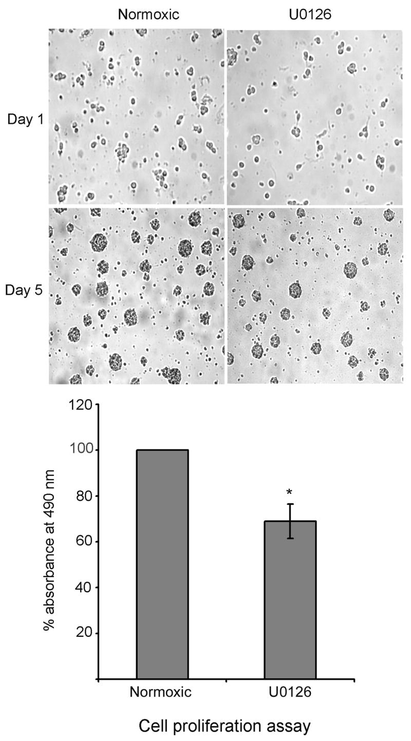
The neurogenic regions such as subventricular zone of the lateral ventricles could become ischemic in some clinical situations due to the blockage of blood vessels by blood clots. Hence the aim of this study is to investigate the effects of OGD on the growth of neural progenitor cells and the phosphorylation of ERK, which plays an important role in the growth of these cells. Oxygen glucose deprivation (OGD) for 4h decreased the growth of neural progenitor cells in vitro and also decreased the phosphorylation of extracellular signal regulated kinase (ERK). Inhibition of the ERK pathway for 4h using U0126 (10 microM) also decreased the growth of progenitor cells. These data suggest that a decline in the phospho-ERK content might decrease the growth of progenitor cells following OGD.


Figures

Fig. 1
Fig. 1
Effect of OGD on the proliferation of neural progenitor cells: Neural progenitor cells were subjected to OGD for 4 h, followed by culturing in neurobasal medium containing B27 and FGF2 for another 5 days. At the end of experimental period, cell proliferation assay was performed to detect the metabolic activity of the cells. The decrease in the metabolic activity of the cells is represented by bar graph. The cells were photographed before and after the experiment. * P < 0.05 as compared to normoxic cells.
Fig. 2
Fig. 2
Effect of OGD on the phosphorylation of ERK. A: Neural progenitor cells were subjected to OGD for 4 h followed by lysis in lysis buffer. The lysate was subjected to immunoblotting using phospho-ERK, ERK and actin antibodies. Note: OGD decreased the phosphorylation of ERK but did not alter the content of ERK. * P < 0.05 as compared to normoxic cells. The samples are shown in triplicate. B: Neural progenitor cells were subjected to OGD for various time points and the cell lysate was processed as described in Fig 2A. * P < 0.05 as compared to 0h incubation. C: Effect of glucose and oxygen on the phosphorylation of ERK. Neural progenitor cells were subjected to OGD, or glucose deprivation or oxygen deprivation for 4 h and the phospho-ERK content was detected as described in Fig. 2A. Note: Oxygen but not glucose decreased the phosphorylation of ERK as compared to normoxic cells.
Fig. 3
Fig. 3
Inhibition of ERK pathway decreases the proliferation of neural progenitor cells. Neural progenitor cells were incubated under normoxic conditions in the presence and absence of U0125 (10uM) (ERK inhibitor) for 4 h. After the experimental period, cells were subsequently cultured in neurobasal medium containing B27 and FGF2 for 5 days. At the end of experimental period the cells were photographed and cell proliferation assay was performed as described in methods. * P < 0.05 as compared to normoxic cells.

References

    1. Palmer TD, Ray J, Gage FH. FGF-2-Responsive Neuronal Progenitors Reside in Proliferative and Quiescent Regions of the Adult Rodent Brain. Mol Cell Neurosci. 1995;6(5):474–486. - PubMed
    1. Martens DJ, Seaberg RM, vander Kooy D. In vivo infusions of exogenous growth factors into the fourth ventricle of the adult mouse brain increase the proliferation of neural progenitors around the fourth ventricle and the central canal of the spinal cord. Eur J Neurosci. 2002;16(6):1045–57. - PubMed
    1. Aberg MA, Aberg ND, Hedbacker H, Oscarsson J, Eriksson PS. Peripheral infusion of IGF-1 selectively induces neurogenesis in the adult rat hippocampus. J Neurosci. 2000;20(8):2896–2903. - PMC - PubMed
    1. Schanzer A, Wachs FP, Wilhelm D, Acker T, Cooper-Kuhn C, Beck H. Direct stimulation of adult neural stem cells in vitro and neurogenesis in vivo by vascular endothelial growth factor. Brain Pathol. 2004;14(3):237–248. - PMC - PubMed
    1. Yamamoto S, Yamamoto N, Kitamura T, Nakamura K, Nakafuku M. Proliferation of parenchymal neural progenitors in response to injury in the adult rat spinal cord. Exp Neurol. 2001;172(1):115–127. - PubMed

Publication types

Substances