

Contrast-enhanced endoscopic ultrasound in discrimination between benign and malignant mediastinal and abdominal lymph nodes
- PMID: 17891499
- PMCID: PMC12161630
- DOI: 10.1007/s00432-007-0309-7
Contrast-enhanced endoscopic ultrasound in discrimination between benign and malignant mediastinal and abdominal lymph nodes
Abstract
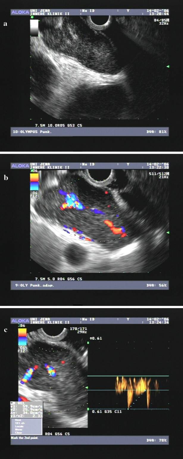
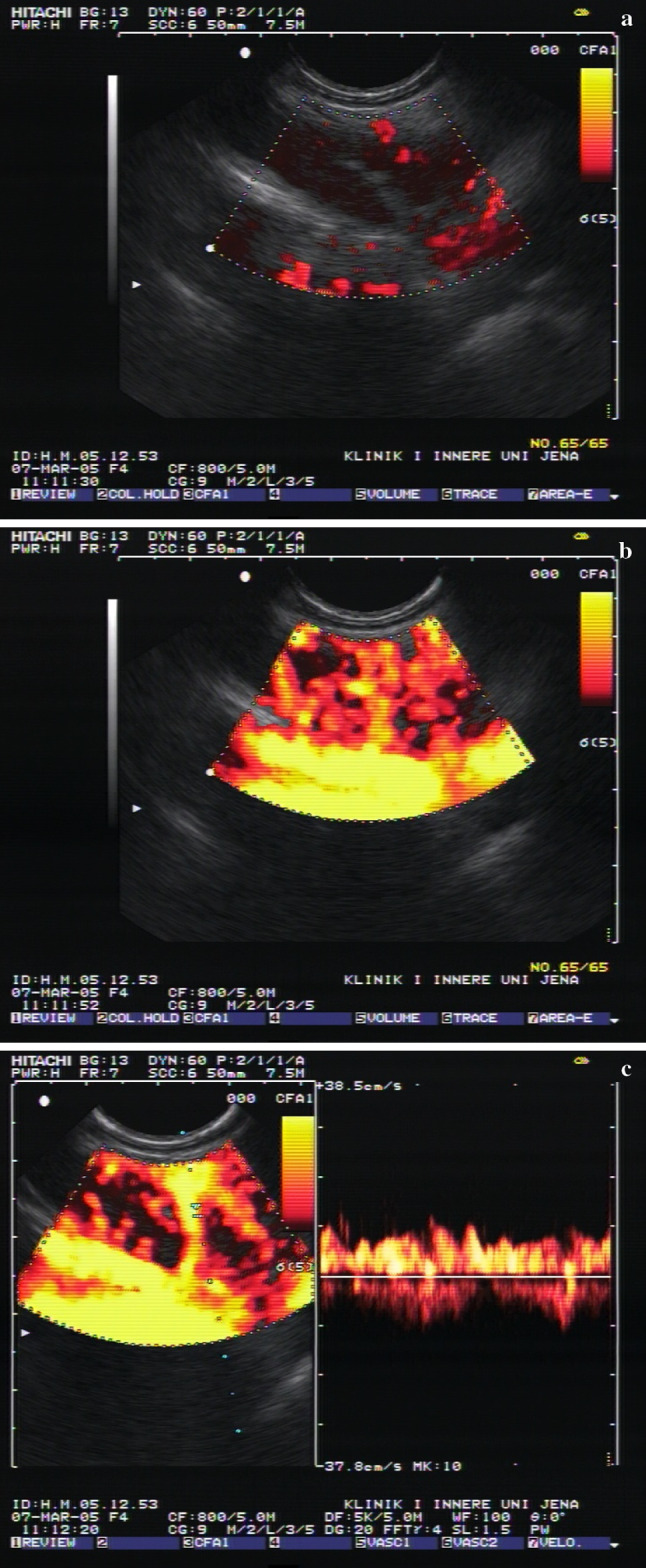
Background: Enlarged lymph nodes in the mediastinum reflect neoplastic, infectious or other diseases. The classification of these nodes is crucial in the management of the patient. Currently, only invasive measures obtaining tissue samples reach satisfying specificity. Contrast-enhanced endoscopic ultrasound (EUS) may offer a non-invasive alternative.
Materials and methods: A total of 122 patients (age: 63 +/- 15 years, 92 males, 30 females) with enlarged mediastinal and/or paraaortic lymph nodes diagnosed by CT scan were included in the study. EUS-guided fine needle aspiration was performed and cytologic specimens were diagnosed as representing a malignant or benign process in case of Papanicolau IV and V, or Papanicolau I and II, respectively.
Results: Based on cytology results, the investigated lymph nodes were classified as neoplastic (n = 48) or non-neoplastic lymph nodes. Using the B-mode criteria the preliminary diagnosis was confirmed in 64 out of 74 benign lymph nodes (specificity 86%). Regarding malignant lymph nodes 33 of 48 were confirmed (sensitivity 68%). Using the advanced contrast-enhanced EUS criteria the diagnosis was confirmed in 68 of 74 benign lymph nodes (specificity 91%). However, in case of malignant lymph nodes the number of correct diagnoses dropped to 29 of 48 lymph nodes (sensitivity 60%). The contrast-enhanced EUS criteria to identify benign lymph nodes and node enlargement in malignant lymphoma do not differ. If those ten patients with malignant lymphoma are excluded, the sensitivity of the contrast enhanced EUS for malignant lymph nodes rises to 73%.
Conclusion: Contrast-enhanced EUS improves the specificity in diagnosing benign lymph nodes as compared to B-mode EUS. It does not improve the correct identification of malignant lymph nodes and cannot replace EUS-guided fine-needle aspiration.
Figures

References
-
- Ahuja A, Ying M (2002) An overview of neck node sonography. Invest Radiol 37:333–342 - PubMed
-
- Baatenburg de Jong RJ, Rongen RJ, Lameris JS, Harthoom M, Verwoerd CD, Knegt P (1989) Metastatic neck disease. Palpation vs. ultrasound examination. Arch Otolaryngol Head Neck Surg 115:689–690 - PubMed
-
- Becker D, Strobel D, Bernatik T, Hahn EG (2001) Echo-enhanced color- and power-Doppler EUS for the discrimination between focal pancreatitis and pancreatic carcinoma. Gastrointest Endosc 53:784–789 - PubMed
-
- Catalano MF, Sivak MV Jr, Rice T, Gragg LA, Van Dam J (1994) Endosonographic features predictive of lymph node metastasis. Gastrointest Endosc 40:442–446 - PubMed
-
- Dietrich CF, Schuesslerm G, Trojan J, Fellbaum C, Ignee A (2005) Differentiation of focal nodular hyperplasia and hepatocellular adenoma by contrast-enhanced ultrasound. Br J Radiol 78:704–707 - PubMed
MeSH terms
Substances
LinkOut - more resources
Full Text Sources
