Review
. 2007;51(6-7):663-78.
doi: 10.1387/ijdb.072367bf.

Molecular evolution of the vertebrate mechanosensory cell and ear

Affiliations
Review

Molecular evolution of the vertebrate mechanosensory cell and ear

Bernd Fritzsch et al. Int J Dev Biol. 2007.

Abstract

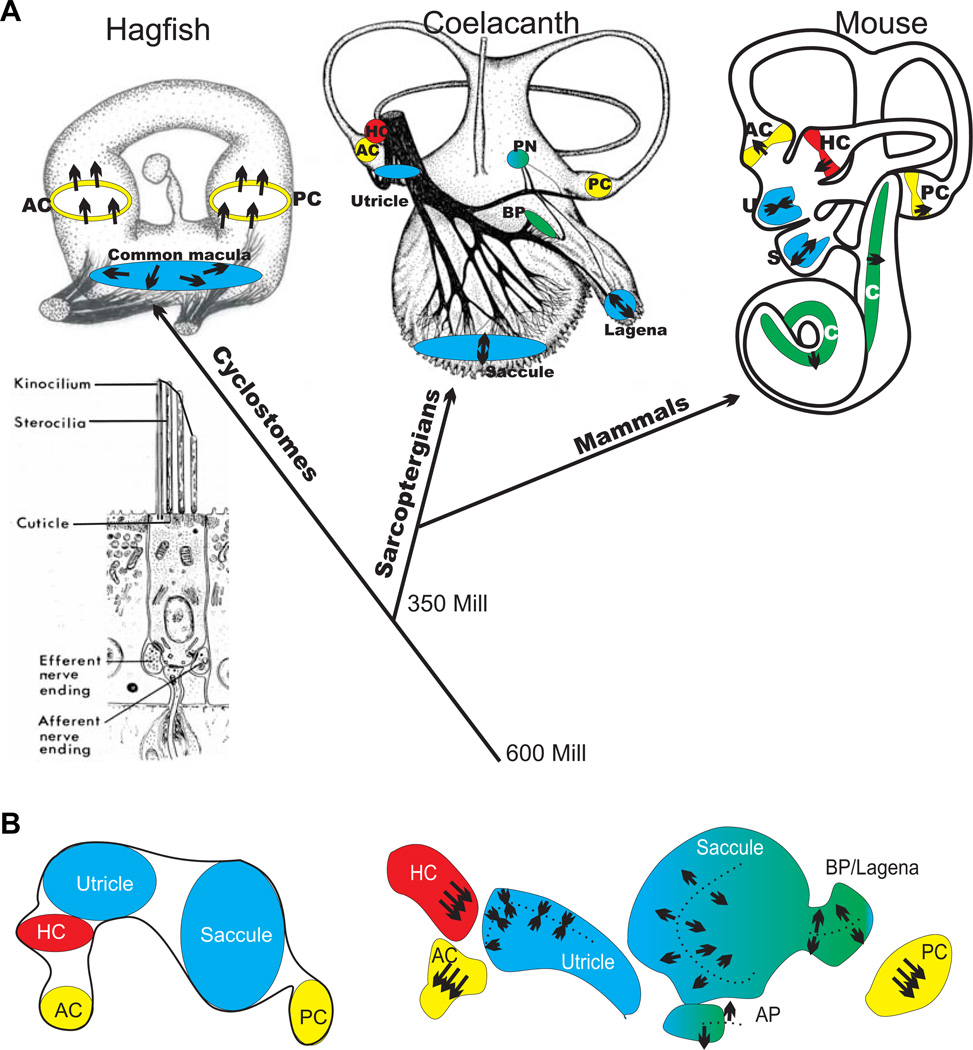
The molecular basis of mechanosensation, mechanosensory cell development and mechanosensory organ development is reviewed with an emphasis on its evolution. In contrast to eye evolution and development, which apparently modified a genetic program through intercalation of genes between the master control genes on the top (Pax6, Eya1, Six1) of the hierarchy and the structural genes (rhodopsin) at the bottom, the as yet molecularly unknown mechanosensory channel precludes such a firm conclusion for mechanosensors. However, recent years have seen the identification of several structural genes which are involved in mechanosensory tethering and several transcription factors controlling mechanosensory cell and organ development; these warrant the interpretation of available data in very much the same fashion as for eye evolution: molecular homology combined with potential morphological parallelism. This assertion of molecular homology is strongly supported by recent findings of a highly conserved set of microRNAs that appear to be associated with mechanosensory cell development across phyla. The conservation of transcription factors and their regulators fits very well to the known or presumed mechanosensory specializations which can be mostly grouped as variations of a common cellular theme. Given the widespread distribution of the molecular ability to form mechanosensory cells, it comes as no surprise that structurally different mechanosensory organs evolved in different phyla, presenting a variation of a common theme specified by a conserved set of transcription factors in their cellular development. Within vertebrates and arthropods, some mechanosensory organs evolved into auditory organs, greatly increasing sensitivity to sound through modifications of accessory structures to direct sound to the specific sensory epithelia. However, while great attention has been paid to the evolution of these accessory structures in vertebrate fossils, comparatively less attention has been spent on the evolution of the inner ear and the central auditory system. Recent advances in our molecular understanding of ear and brain development provide novel avenues to this neglected aspect of auditory neurosensory evolution.


Figures

Fig. 1
Fig. 1. Various channels that respond to changes in turgor resulting in stretch of the membranes (double arrow in A, B) have been found in single cell organisms
Detailed models of the pentameric mechanosensitive channel of bacteria suggests an iris-like opening upon tension acting in the plane of the membrane (B). It is possible that such mechanosensitive channels were modified in the unicellular ancestor of metazoans through extracellular or intracellular matrix attachments to provide increased sensitivity for shearing forces. Molecular evidence suggests that, across metazoans, only members of two cation channel families are candidates for mechanosensitive channels (C,D). It thus remains possible that metazoan ancestors evolved either or both families for specific properties that allow increased sensitivity to detect mechanical stimulations. What such properties could be remains unknown in the absence of any model of sensitive mechanosensory channel in any metazoan taxon. Mutational analysis has identified several genes that are essential for the function in nematodes (C) and vertebrates (D). In nematodes, fine touch is lost when either specific molecules of the extracellular matrix to which the channel is anchored are lost (Mec-1, 5, 9), or if specific components of the channel complex are lost (Mec-2, 4, 10, 14). However, loss to the intracellular tubules (Mec-7, 12) may lead only to a reduced sensitivity, not a complete loss of sensation. In contrast, in vertebrates there is no extracellular matrix or cuticular connection. Instead, two stereocilia are interconnected presumably by Cdh23 that is hypothesized to be anchored to MyoVIIa via harmonin. Loss of any of these genes results in deafness indicating that in vertebrates mechanosensation requires relative movement against the actin core of the stereocilia. Additional connections exist between Cdh23 and Myo1c but no knockout data support the claimed function as an adaptor. It is speculated that MyoVIIa transports the still unknown amiloride sensitive mechanosensory channel to the tip but it is unclear whether this connection remains past development. In nematodes at least two essential subunits of the mechanosensitive channel are known whereas it is not clear what the vertebrate channel is composed of. Certain candidates have been excluded as mutants in, for example, TRPA1 do hear excluding an essential role of this protein in mechanosensory transduction. Note that both nematode and vertebrate have a shaker-type channel associated with the mechanosensory channel, but details are unknown. Modified after (Bryant et al., 2005, Chiang et al., 2004, Syntichaki and Tavernarakis, 2004).
Fig. 2
Fig. 2. The evolution of the atonal and neurogenin families of bHLH genes (A) and the evolution of the mechanosensory cells and their associated neurons that require those family members for cellular development (B) is shown
Note that atonal and achaete/scute family evolved already in coelenterates. However, the neurogenin family may have evolved only in triploblasts. Evolution of a pair of cells (a secondary mechanosensory cell without an axon and a mechanosensory neuron connecting the cell to the brain) evolved out of a primary mechanosensory cell (with an axon) only after the neurogenin family had evolved. Whether atonal family members are always associated with mechanosensory cells in triploblasts and whether neurons associated with mechanosensory cells in other triploblasts require neurogenin for development is unknown. Modified after (Fritzsch and Beisel, 2004, Furlong and Graham, 2005).
Fig. 3
Fig. 3. Evolution of mechanosensory cells
Kinocilia (red) and microvilli (light blue) of known or suspected mechanosensory cells in various eukaryotic unicellular (1) and multicellular (3) organisms are shown. Orthologues of structural genes relevant for mechanosensation or for development of polarity such as actin, tubulin, rare myosins, cadherins, espin, β- catenin and Wnt genes and several transcription factors are known in protists, Diploblasts (2) and various triploblasts and are thus ancestral to vertebrates. Note that the single celled ancestor of all multicellular animals, the choanoflagellates (1), has a single, actively beating kinocilium surrounded by microvilli that carry an actin core (A). In some diploblasts the central kinocilium is surrounded by an asymmetric assembly of microvilli of various diameters, potentially providing directional sensitivity (B). Among deuterostomes, urochordates have various presumed mechanosensory cells that have a kinocilium with asymmetrically arranged microvilli. Vertebrates are unique in that a highly polarized, organ-pipe assembly of actin rich stereocilia is attached via tip links with each other and the asymmetrically placed kinocilium. Among protostomes, mollusks may have numerous, interconnected kinocilia on mechanosensory cells. Ecdysozoans have either mechanosensory cells with cilia or have a kinocilium that is stretched by the stimulus. Arrows indicate the direction of stimulation. Statocysts are known for many taxa of metazoans, but a lateral line system is restricted to few. Modified after (Arkett et al., 1988, Budelmann, 1989, Burighel et al., 2003, Fritzsch et al., 2006b, Jorgensen, 1989, Steenkamp et al., 2006, Todi et al., 2005).
Fig. 4
Fig. 4. Comparison of the genes involved in trachea formation in flies with inner ear development in mice
Icons of the same shape and color represent homologous genes in Drosophila m. and Mus m. Known interactions between genes are demonstrated by the black arrows. Several homologous genes have been identified in these developmental pathways, but their interactions have not yet been fully described in either species. Putative sequence of activation is from the top with the fly and louse Fgf/Fgfr system providing the integration between patterning events outside the trachea/ear and intracellular signaling that leads to morphogenesis The striking resemblance of these genes as a complex interacting module supports a model in which not only are individual genes conserved across phyla, but the entire signaling network is conserved and utilized in the development of diverse structures, governing various aspects of branching morphogenesis. We recently tested the predictive value of this model and reported on the effect of Foxg1, the orthologue of Slp1 (Lee and Frasch, 2004, Mondal et al., 2007), in mouse ear development (Pauley et al., 2006). More of the listed genes should be tested for such conserved function to assess how much of the entire developmental module is invariable and thus needs to be inherited as an entire cascade of gene interactions with modifications to suit the need of the specific tissue in question. Modified after (Swantek and Gergen, 2004). Gene names follow the nomenclature as published in PubMed.
Fig. 5
Fig. 5. Crucial steps in vertebrate ear evolution (A) and development (B) are depicted
It is assumed that the vertebrate hair cells with afferents and efferents co-evolved with the ear (insert) some 600 million years ago. Note that the ears of the three depicted vertebrate species differ in the number of canal cristae (hagfish has two, coelacanth [Latimeria] and mouse have three), number of vestibular organs (hagfish has one [common macula], coelacanth has three [utricle, U; saccule, S; lagena, L] and mouse has two [utricle, U; saccule, S]) and number of organs near perilymphatic ducts (none in hagfish, two [basilar papilla, BP; papilla neglecta, PN] in coelacanth and one [organ of Corti of the cochlea, C] in mouse). Major morphological evolutionary changes are the addition of a horizontal canal in gnathostomes and the transformation of the utricle into several recesses containing the saccule, lagena and cochlea. It is suggested that the evolution of up to nine sensory organs of the vertebrate ear (A) comes about through ontogenetic segregation of a single primordium into multiple sensory patches (B). After segregation, each sensory patch differentiates along a unique trajectory to form adult epithelia that perceive discrete aspects of the mechanical stimulation that reaches the ear (A). Development (B) therefore recapitulates the evolutionary segregation and differentiation of various epithelia from a common precursor. Integrated into this differentiation is the organization of different polarities of hair cells (arrows in B) that can be opposing (utricle, saccule, lagena) or one polarity (canal cristae). Note that the polarity of hair cells in the cristae is similar in anterior and posterior crista (away from the gravistatic organs), whereas the horizontal crista is polarized toward the gravistatic organs. How the ancestral molecular pathway to set up cellular polarity in the various sensory epithelia has been modified remains unclear. Modified after (Fritzsch and Beisel, 2004, Fritzsch et al., 2002).
Fig. 6
Fig. 6. The transformation of the ear, sensory neurons and brainstem from a non-auditory, primitive condition, into the derived condition, enabling a tetrapod vertebrate to hear, is shown
The primitive ear has vestibular sensory epithelia (VE) that are connected with vestibular sensory neurons (VN) to the vestibular nuclei of the brainstem (VeNu). Additional sensory systems in primary aquatic vertebrates are the electroreceptive ampullary organs (AO) and the mechanosensory neuromasts of the lateral line (NEU). These organs are connected via specific sets of sensory neurons (ELL, LL) to specific brainstem nuclei (ELLNu, LLNu). Derived land vertebrates have lost these senses and have a sound pressure receiving sense, called hearing. This sense is characterized by the auditory epithelium (AE) that sits at or near a sound conducting perilymphatic system (PLS) and is covered with a tectorial membrane (TM). Auditory neurons (AN) conduct the information from the auditory epithelium to the auditory nuclei (AuNu) of the brainstem. This basic organization may have evolved in the aquatic ancestor of terrestrial vertebrates, but was modified in amphibians through the addition of the amphibian papilla and in amniotes through the formation of the cochlea. Modified after (Fritzsch and Neary, 1998).

References

    1. Alonso CR, Wilkins AS. The molecular elements that underlie developmental evolution. Nat Rev Genet. 2005;6:709–715. - PubMed
    1. Alvarez Y, Alonso MT, Vendrell V, Zelarayan LC, Chamero P, Theil T, Bosl MR, Kato S, Maconochie M, Riethmacher D, et al. Requirements for FGF3 and FGF10 during inner ear formation. Development. 2003;130:6329–6338. - PubMed
    1. Arendt D, Tessmar-Raible K, Snyman H, Dorresteijn AW, Wittbrodt J. Ciliary photoreceptors with a vertebrate-type opsin in an invertebrate brain. Science. 2004;306:869–871. - PubMed
    1. Arkett SA, Mackie GO, Meech RW. Hair cell mechanoreception in a jellyfish Aglantha digitale. Journal of Experimental Biology. 1988;135:329–342.
    1. Bailey AP, Bhattacharyya S, Bronner-Fraser M, Streit A. Lens specification is the ground state of all sensory placodes, from which FGF promotes olfactory identity. Dev Cell. 2006;11:505–517. - PubMed

Publication types