. 2007 Sep 21;3(9):1240-9.
doi: 10.1371/journal.ppat.0030127.

Immunodominant tuberculosis CD8 antigens preferentially restricted by HLA-B

Affiliations

Immunodominant tuberculosis CD8 antigens preferentially restricted by HLA-B

Deborah A Lewinsohn et al. PLoS Pathog. .

Abstract

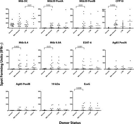
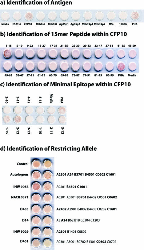
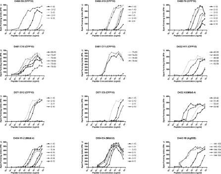
CD8(+) T cells are essential for host defense to intracellular bacterial pathogens such as Mycobacterium tuberculosis (Mtb), Salmonella species, and Listeria monocytogenes, yet the repertoire and dominance pattern of human CD8 antigens for these pathogens remains poorly characterized. Tuberculosis (TB), the disease caused by Mtb infection, remains one of the leading causes of infectious morbidity and mortality worldwide and is the most frequent opportunistic infection in individuals with HIV/AIDS. Therefore, we undertook this study to define immunodominant CD8 Mtb antigens. First, using IFN-gamma ELISPOT and synthetic peptide arrays as a source of antigen, we measured ex vivo frequencies of CD8(+) T cells recognizing known immunodominant CD4(+) T cell antigens in persons with latent tuberculosis infection. In addition, limiting dilution was used to generate panels of Mtb-specific T cell clones. Using the peptide arrays, we identified the antigenic specificity of the majority of T cell clones, defining several new epitopes. In all cases, peptide representing the minimal epitope bound to the major histocompatibility complex (MHC)-restricting allele with high affinity, and in all but one case the restricting allele was an HLA-B allele. Furthermore, individuals from whom the T cell clone was isolated harbored high ex vivo frequency CD8(+) T cell responses specific for the epitope, and in individuals tested, the epitope represented the single immunodominant response within the CD8 antigen. We conclude that Mtb-specific CD8(+) T cells are found in high frequency in infected individuals and are restricted predominantly by HLA-B alleles, and that synthetic peptide arrays can be used to define epitope specificities without prior bias as to MHC binding affinity. These findings provide an improved understanding of immunodominance in humans and may contribute to a development of an effective TB vaccine and improved immunodiagnostics.


Conflict of interest statement

Competing interests. The authors have declared that no competing interests exist.

Figures

Figure 1
Figure 1. Determination of Human Effector Cell Frequencies Ex Vivo Using the IFN-γ ELISPOT Assay
Magnetic bead–purified CD8+ T cells from a single donor (D466) were cultured with DCs (20,000/well) either infected with Mtb (H37Rv, multiplicity of infection = 50) or pulsed with peptide pool representing CFP10 (5 μg/ml each peptide; 15-mer overlap by 11 aa) in an IFN-γ ELISPOT assay. Each responding T cell population was tested in duplicate at four different cell concentrations. To determine the effector cell frequency of antigen-specific T cells, the average number of spot forming units per well for each duplicate was plotted against the number of responder cells per well. Linear regression analysis was used to determine the slope of the line, which represents the frequency of antigen-specific T cells. The assay was considered positive, reflecting the presence of a primed T cell response, if the binomial probability for the number of spots was significantly different by experimental and control assays, i.e., if the experimental line is statistically significantly different from the control line. The frequency of Mtb-specific and CFP10-specific T cells demonstrated in this figure was 1/307 and 1/1676, respectively.
Figure 2
Figure 2. High Ex vivo CD8+ T Cell Frequencies to Mtb Antigens Are Associated with Mtb Infection
As in Figure 1, to determine ex vivo CD8+ T cell frequencies, autologous DCs either infected with Mtb (graph labeled “Mtb DC”) or pulsed with cognate peptide pools representing known Mtb CD4 antigens (all other graphs) were incubated with CD8+ T cells in an IFN-γ ELISPOT assay. The number of spot forming units (SFU)/250,000 cells is shown, a number which is derived from linear regression analysis of the frequency of T cells tested at concentrations of 5 × 10⁵, 2.5 × 10⁵, 1.2 × 10⁵, and 6 × 10⁴ cells/well. Individuals without evidence for Mtb infection (“normal”); those with LTBI, and those with active TB (“active” defined as culture confirmed pulmonary TB) were evaluated. “Mtb Infected” includes both the “LTBI” and “active” groups. The number of individuals tested for each antigen were: Mtb infection (normal, n = 11; LTBI, n = 11; active, n = 6); Mtb39 Pool A (normal, n = 14; LTBI, n = 20; active, n = 10); Mtb39 Pool B (normal, n = 14; LTBI, n = 20; active, n = 10); CFP10 (normal, n = 14; LTBI, n = 20; active, n = 10); Mtb8.4 (normal, n = 14; LTBI, n = 20; active, n = 10); Mtb9.9A (normal, n = 14; LTBI, n = 20; active, n = 5); ESAT-6 (normal, n = 14; LTBI, n = 20; active, n = 5); Ag85 Pool A (normal, n = 14; LTBI, n = 17; active, n = 5); Ag85 Pool B (normal, n = 14; LTBI, n = 17; active, n = 5); 19kDa (normal, n = 12; LTBI, n = 17; active, n = 5); and EsxG (normal, n = 14; LTBI, n = 17; active, n = 5). p-Values are noted where statistically significant differences between groups are noted (p = <0.05; Wilcoxon/Kruskal–Wallis).
Figure 3
Figure 3. Definition of Antigenic Specificity and HLA Restriction: Characterization of T Cell Clone D466 D6
(A–C) To identify the antigen and minimal epitope recognized by T cell clone D466 D6, T cells (5,000 cells/well) were incubated with autologous LCL (20,000/well) and antigen (5 μg/ml). Negative controls (media, no antigen) and positive controls (phytohemagglutanin [PHA], final, 10 μg/ml) are shown for each assay. IFN-γ was assessed by ELISPOT after 18 h of co-culture. Pictures of ELISPOT wells are shown. (A) Antigens consisted of peptide pools representing known CD4 Mtb antigens, made up of 15-aa peptides overlapping by 11 aa. (B) Antigens consisted of individual 15-aa CFP10 peptides that together constitute the peptide pool. (C) Antigens consisted of individual nested peptides within CFP101–15 (10 aa, 9 aa, or 8 aa), used to further map the epitope. (D) The restricting allele was identified using LCLs (20,000/well) that were either autologous or expressing HLA alleles matching D466 at one or two alleles (shown in bold font), pulsed with CFP102–10 (5 μg/ml) as APC. Autologous LCLs without peptide (control) are also shown. After 2 h, cells were washed and incubated with T cells (500 cells/well) in an IFN- γ ELISPOT assay. Pictures of ELISPOT wells are shown.
Figure 4
Figure 4. Confirmation of Minimal Epitope Mapping of D466 D6
To confirm the minimal epitope, autologous LCLs (20,000/well) were pulsed with peptide at the concentration indicated and co-cultured with T cells (1,000 cells/well). IFN-γ was assessed by ELISPOT after 18 h co-culture. Each point represents the mean of duplicate determinations.
Figure 5
Figure 5. Summary of Minimal Epitope Mapping Data
To determine the minimal epitope, autologous LCLs (20,000/well) were pulsed with peptide at the concentration indicated and co-cultured with T cells (1,000 cells/well). IFN-γ was assessed by ELISPOT after 18 h co-culture. Each point represents the mean of duplicate determinations.
Figure 6
Figure 6. Profiling of Immunodominance Pattern for CFP10
To determine the effector cell frequencies, autologous DCs (20,000/well) were pulsed either with each individual 15-mer peptide (5 μg/ml), the peptide pool (PP; 5 μg/each peptide), or the minimal epitope (ME) determined from T cell clones derived from each donor (D466:CFP102–11; D480:CFP103–11; D481:CFP1075–83; 5 μg/ml), and tested against 250,000 magnetic bead–purified CD8+ T cells. IFN-γ release was assessed by ELISPOT after 18 h of co-culture. Each point represents the mean of duplicate determinations.

References

    1. WHO report 2007. Geneva: World Health Organization; 2007. Global tuberculosis control: Surveillance, planning, financing.
    1. The global plan to stop TB, 2006–2015 / Stop TB Partnership. Geneva: World Health Organization; 2006.
    1. Flynn JL, Chan J. Immunology of tuberculosis. Annu Rev Immunol. 2001;19:93–129. - PubMed
    1. North RJ, Jung YJ. Immunity to tuberculosis. Annu Rev Immunol. 2004;22:599–623. - PubMed
    1. Oldstone MB. The role of cytotoxic T lymphocytes in infectious disease: History, criteria, and state of the art. Curr Top Microbiol Immunol. 1994;189:1–8. - PubMed

Publication types

MeSH terms