. 2007 Nov;150(2):358-67.
doi: 10.1111/j.1365-2249.2007.03495.x. Epub 2007 Sep 24.

Hypoxia attenuates effector-target cell interaction in the airway and pulmonary vascular compartment

Affiliations

Hypoxia attenuates effector-target cell interaction in the airway and pulmonary vascular compartment

S Meyer et al. Clin Exp Immunol. 2007 Nov.

Abstract

Leucocyte infiltration is known to play an important role in hypoxia-induced tissue damage. However, little information is available about hypoxia and interaction of effector (neutrophils) with target cells (alveolar epithelial cells, AEC; rat pulmonary artery endothelial cells, RPAEC). The goal of this study was to elucidate hypoxia-induced changes of effector-target cell interaction. AEC and RPAEC were exposed to 5% oxygen for 2-6 h. Intercellular adhesion molecule-1 (ICAM-1) expression was determined and cell adherence as well as cytotoxicity assays were performed. Nitric oxide and heat shock protein 70 (HSP70) production was assessed in target cells. Under hypoxic conditions enhanced ICAM-1 production was found in both cell types. This resulted in an increase of adherent neutrophils to AEC and RPAEC. The death rate of hypoxia-exposed target cells decreased significantly in comparison to control cells. Nitric oxide (NO) concentration was enhanced, as was production of HSP70 in AEC. Blocking NO production in target cells resulted in increased cytotoxicity in AEC and RPAEC. This study shows for the first time that target cells are more resistant to effector cells under hypoxia, suggesting hypoxia-induced cell protection. An underlying mechanism for this phenomenon might be the protective effect of increased levels of NO in target cells.


Figures
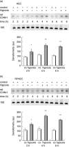
Fig. 1
Fig. 1
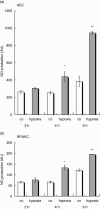
Intercellular adhesion molecule-1 (ICAM-1) expression in alveolar epithelial cells (AEC) (a) and rat pulmonary artery endothelial cells (RPAEC) (b). AEC or RPAEC were exposed to 5% oxygen (hypoxia) or 21% oxygen (control, co) for 2, 4 and 6 h. RNA was extracted and reverse transcriptase–polymerase chain reaction was performed. Densitometry values are shown as means ± standard error of the mean from five different experiments. AEC: *P < 0·05, **P < 0·001; RPAEC: *P < 0·01, **P < 0·001.
Fig. 2
Fig. 2
Neutrophil adhesion to alveolar epithelial cells (AEC) (a) and rat pulmonary artery endothelial cells (RPAEC) (b), exposed previously to 5% oxygen (hypoxia) or 21% oxygen (control, co). Neutrophils (2 × 10⁵/well) were added to AEC or RPAEC for 30 min. Non-adherent cells were washed away and adherent cells were counted. Values are shown as means ± standard error of the mean from five different experiments. AEC: *P < 0·001, **P < 0·0005; RPAEC: *P < 0·01, **P < 0·0005.
Fig. 3
Fig. 3
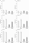
Alveolar epithelial cell (AEC) (a) and rat pulmonary artery endothelial cell (RPAEC) (b) killing by activated neutrophils. Cytotoxicity assay was performed by measuring lactate dehydrogenase (LDH) release. Neutrophils (2 × 10⁵/well) were added to AEC or RPAEC, stimulated with phorbol 12-myristate 13-acetate and co-incubated with target cells in 5% oxygen (hypoxia) or 21% oxygen (control, co) for 2, 4 and 6 h. Cytotoxicity (%) was calculated based on total content and spontaneous release of LDH. Values are shown as means ± standard error of the mean from five different experiments. AEC: *P < 0·05, **P < 0·01; RPAEC: *P < 0·005, **P < 0·0005.
Fig. 4
Fig. 4
Determination of intracellular nitric oxide (NO) production in alveolar epithelial cells (AEC) (a) and rat pulmonary artery endothelial cells (RPAEC) (b). Cells were exposed to 5% oxygen (hypoxia) or 21% oxygen (control) for 2, 4 and 6 h, and were loaded with 10 μM of the membrane-permeable 4,5-diaminofluorescein-2/diacetate (DAF-2/DA) for 30 min. After washing the cells once with 1% Dulbecco's modified Eagle's medium fluorescence was measured at 485 nm (excitation) and at 535 nm (emission) (values as arbitrary unit; au). Values are shown as means ± standard error of the mean from five different experiments. AEC: *P < 0·05, **P < 0·01; RPAEC: *P < 0·05, **P < 0·01.
Fig. 5
Fig. 5
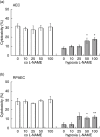
Effect of nitric oxide synthase inhibitor NW-nitro-L-arginine methyl ester (L-NAME) on effector cell-induced alveolar epithelial cell (AEC) (a) and rat pulmonary artery endothelial cell (RPAEC) (b) killing using different concentrations of L-NAME. AEC and RPAEC were pretreated with 10, 25, 50 or 100 μM L-NAME for 30 min. Neutrophils (2 × 10⁵/well) were added to AEC or RPAEC, stimulated with phorbol 12-myristate 13-acetate and co-incubated with target cells in 5% oxygen (hypoxia) or 21% oxygen (control, co) for 4 h. Cytotoxicity (%) was calculated based on total content and spontaneous release of lactate dehydrogenase (LDH). Values are shown as means ± standard error of the mean from five different experiments. AEC: *P < 0·05; RPAEC: *P < 0·05, **P < 0·001.
Fig. 6
Fig. 6
Effect of nitric oxide synthase inhibitor NW-nitro-L-arginine methyl ester (L-NAME) on effector cell-induced alveolar epithelial cell (AEC) (a) and rat pulmonary artery endothelial cell (RPAEC) (b) killing at 2, 4 and 6 h of hypoxia/normoxia. AEC and RPAEC were pretreated with 50 μM L-NAME for 30 min. Neutrophils (2 × 10⁵/well) were added to AEC or RPAEC, stimulated with phorbol 12-myristate 13-acetate and co-incubated with target cells in 5% oxygen (hypoxia) or 21% oxygen (control, co) for 2, 4 or 6 h. Cytotoxicity (%) was calculated based on total content and spontaneous release of lactate dehydrogenase (LDH). Values are shown as means ± standard error of the mean from five different experiments. AEC: *P < 0·05, **P < 0·005; RPAEC: *P < 0·05, **P < 0·01.
Fig. 7
Fig. 7
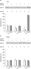
Expression of heat shock protein 70 (HSP70) in alveolar epithelial cell (AEC) (a) and rat pulmonary artery endothelial cells (RPAEC) (b) after exposure to 5% oxygen (hypoxia) or 21% oxygen (control, co) for 2, 4 and 6 h. Cells were lysed and Western blot analysis was performed (one blot shown). Values are shown as means ± standard error of the mean from three different experiments. AEC: *P < 0·01.

References

    1. Beck-Schimmer B, Schimmer RC, Madjdpour C, Bonvini JM, Pasch T, Ward PA. Hypoxia mediates increased neutrophil and macrophage adhesiveness to alveolar epithelial cells. Am J Respir Cell Mol Biol. 2001;25:780–7. - PubMed
    1. Andonegui G, Bonder CS, Green F, et al. Endothelium-derived Toll-like receptor-4 is the key molecule in LPS-induced neutrophil sequestration into lungs. J Clin Invest. 2003;111:1011–20. - PMC - PubMed
    1. Kuo HP, Lin HC, Hwang KH, Wang CH, Lu LC. Lipopolysaccharide enhances substance P-mediated neutrophil adherence to epithelial cells and cytokine release. Am J Respir Crit Care Med. 2000;162:1891–7. - PubMed
    1. Reutershan J, Ley K. Bench-to-bedside review: acute respiratory distress syndrome − how neutrophils migrate into the lung. Crit Care. 2004;8:453–61. - PMC - PubMed
    1. Beck-Schimmer B, Schimmer RC, Warner RL, et al. Expression of lung vascular and airway ICAM-1 after exposure to bacterial lipopolysaccharide. Am J Respir Cell Mol Biol. 1997;17:344–52. - PubMed

Publication types

MeSH terms