. 2007 Dec;98(12):1914-20.
doi: 10.1111/j.1349-7006.2007.00618.x. Epub 2007 Sep 24.

Downregulation of microRNAs-143 and -145 in B-cell malignancies

Affiliations

Downregulation of microRNAs-143 and -145 in B-cell malignancies

Yukihiro Akao et al. Cancer Sci. 2007 Dec.

Abstract

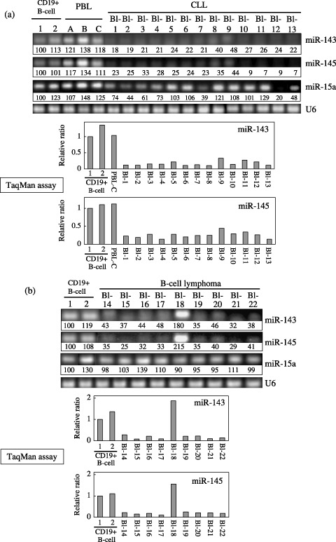
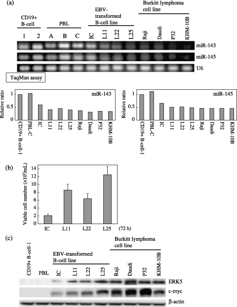
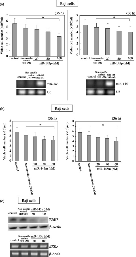
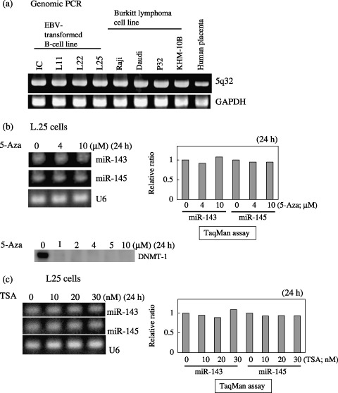
Recently, it has been found that inappropriate expression of microRNAs (miRNAs) is strongly associated with carcinogenesis. In this study, we demonstrated that the expression of miRNAs (miRs) -143 and -145, the levels of which were previously shown to be reduced in colon cancers and various kinds of established cancer cell lines, was also decreased in most of the B-cell malignancies examined, including chronic lymphocytic leukemias (CLL), B-cell lymphomas, Epstein-Barr virus (EBV)-transformed B-cell lines, and Burkitt lymphoma cell lines. All samples from 13 CLL patients and eight of nine B-cell lymphoma ones tested exhibited an extremely low expression of miRs-143 and -145. The expression levels of miRs-143 and -145 were consistently low in human Burkitt lymphoma cell lines and were inversely associated with the cell proliferation observed in the EBV-transformed B-cell lines. Moreover, the introduction of either precursor or mature miR-143 and -145 into Raji cells resulted in a significant growth inhibition that occurred in a dose-dependent manner and the target gene of miRNA-143 was determined to be ERK5, as previously reported in human colon cancer DLD-1 cells. Taken together, these findings suggest that miRs-143 and -145 may be useful as biomarkers that differentiate B-cell malignant cells from normal cells and contribute to carcinogenesis in B-cell malignancies by a newly defined mechanism.


Figures

Figure 1
Figure 1
Expression of microRNAs (miRNAs)‐143 and ‐145 in human B‐cell malignancies examined by quantitative reverse transcription‐polymerase chain reaction (RT‐PCR). Blood samples or lymph nodes (Bl) from patients were obtained on admission or after relapse of the disease. Thirteen cases of chronic lymphocytic leukemia (CLL) (a) and nine cases of B‐cell lymphoma (b) were examined. Peripheral blood lymphocytes (PBL) from individuals a, b, and c and CD19+ B‐cells from the tonsils (1 and 2) were used as controls. In CLLs, the expression of miR‐15a( 28 ) was also examined by semi‐quantitative RT‐PCR. U6 was used as an internal standard. The intensity of the bands for miR‐143, ‐145 and ‐15a expression was determined by densitometry and the value is given under each band. The levels of CD19+ B‐cells for CLL and B‐cell lymphoma were designated as 100 in semi‐quantitative RT‐PCR and as 1 in quantitative RT‐PCR by TaqMan assays using a real‐time PCR. The results of real‐time PCR are expressed as the mean values of two independent experiments.
Figure 2
Figure 2
Expression of microRNAs (miRNAs)‐143 and ‐145 and cell growth in B‐cell cultured cell lines. (a) Evaluation of expression of miRNAs‐143 and ‐145 in human Epstein‐Barr virus (EBV)‐transformed B‐cell lines and Burkitt lymphoma cell lines by use of semi‐quantitative reverse transcription‐polymerase chain reaction (RT‐PCR) and TaqMan MicroRNA assays using real‐time PCR. U6 was used as an internal standard. CD19+ B‐cell‐1 was designated as 1 in quantitative RT‐PCR by TaqMan miRNA assays using real‐time PCR. The results of real‐time PCR were expressed as the mean values of two independent experiments. (b) Steady‐state cell growth of EBV‐transformed B‐cell lines at 72 h after seeding at the concentration of 1 × 105/mL (c) Western blot analysis of ERK5 and c‐myc in control and B‐cell lines in the same samples as in (b), and in human Burkitt lymphoma cell lines. β‐actin was used as an internal control.
Figure 3
Figure 3
Effect of transfection of human Burkitt lymphoma Raji cells with either precursor (a) or mature type (b) of microRNAs (miRNAs)‐143 and ‐145, and ERK5 expression in the cells transfected with the precursor miR‐143 (c). (a, b) Number of viable transfected or control cells at 36 h after transfection is shown. Data are presented as the mean ± standard deviation (SD) of three different experiments, each carried out in duplicate. Levels of miRNAs‐143 and ‐145 in Raji cells at 36 h after the transfection of the cells with miR‐143 or ‐145 precursor miRNAs at 100 nM are shown in (a). U6 was used as an internal standard. The difference between the non‐specific control and 100 (a) or 60 (b) nM treatment was significant (*P < 0.01). (c) Expression levels of ERK5 protein and the mRNA at 36 h after the transfection of Raji cells with miR‐143 precursor miRNAs, as evaluated by western blot analysis (upper panel) and by quantitative RT‐PCR (lower panel), respectively. β‐actin was used as an internal standard. Control cells were incubated in medium containing transfection reagent alone.
Figure 4
Figure 4
Confirmation of the presence of the genomic loci of microRNAs (miRNAs)‐143 and ‐145 at chromosome 5q32 by genomic polymerase chain reaction (PCR) (a) and expression of the miRNAs in Epstein‐Barr virus (EBV)‐transformed L25 cells after 24‐h treatment with 5‐Aza‐2′‐deoxycytidine (5‐Aza, b) or tricostatin A (TSA, c). (a) The primers amplified the DNA fragment including the loci of miRs‐143 and ‐145. Placental DNA was used as a positive control. The genomic locus of glyceraldehyde phosphate dehydrogenase (GAPDH) was used as an internal control. (b, c) The expression of the miRNAs in L25 cells after the treatment with either agent at the indicated concentrations is shown. The value for the control cells is designated as 1 in quantitative reverse transcription (RT)‐PCR by TaqMan miRNA assays using a real‐time PCR. The results of real‐time PCR are expressed as the mean values of two independent experiments. U6 was used as an internal standard. Results of Western blot analysis of DNA methyltransferase (DNMT)‐1 are also given in (b).

References

    1. Lee RC, Feinbaum RL, Ambros V. The C. elegans heterochronic gene lin‐4 encodes small RNAs with antisense complementarity to lin‐14. Cell 1993; 75: 843–54. - PubMed
    1. Lagos‐Quintana M, Rauhut R, Lendeckel W, Tuschl T. Incorporating structure to predict microRNA targets. Science 2001; 294: 853–8. - PubMed
    1. Lau NC, Lim LP, Weinstein EG, Bartel DP. An abundant class of tiny RNAs with probable regulatory roles in Caenorhabditis elegans. Science 2001; 294: 858–62. - PubMed
    1. Lee RC, Ambros V. An extensive class of small RNAs in Caenorhabditis elegans. Science 2001; 294: 862–4. - PubMed
    1. Bartel DP. MicroRNAs: genomics, biogenesis, mechanism, and function. Cell 2004; 116: 281–97. - PubMed

Publication types

MeSH terms