. 2007 Sep 24:5:106.
doi: 10.1186/1477-7819-5-106.

Inflammatory pseudotumor of the Kidney

Affiliations

Inflammatory pseudotumor of the Kidney

David R Selvan et al. World J Surg Oncol. .

Abstract

Background: Inflammatory pseudotumor of the kidney or inflammatory myofibroblastic tumor (IMT) is composed of spindle cells admixed with variable amount of proliferating myofibroblasts, fibroblasts, extracellular collagen, lymphocytes and plasma cells. This mainly affects the urinary bladder or prostate. Renal involvement is rare.

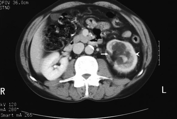
Case presentation: A 56 year-old man was diagnosed with asymptomatic left sided hydronephrosis while being investigated for rheumatoid arthritis. CT scan imaging showed ill defined fascial plains around the kidney and thickening around the renal hilum suggestive of localized inflammatory change. Worsening intermittent left loin pain with increasing hydronephrosis, significant cortical thinning and marked deterioration of renal function necessitated nephrectomy. Macroscopy showed a hydronephrotic fibrotic kidney with microscopy and immunohistochemistry consistent with a histological diagnosis of IMT.

Conclusion: We report a case of an inflammatory pseudotumor of the kidney. It is unique in that the patient presented with painless hydronephrosis followed two years later with progressive deterioration in renal function and worsening loin pain.


Figures

Figure 1
Figure 1
CT scan showing PUJ obstruction (arrow) with ill defined fascial planes around the left kidney (arrow head) that is consistent with inflammation.
Figure 2
Figure 2
Photomicrograph showing a] Vaguely nodular areas of collagenous tissue with scattered spindle cells and darker inflammatory cells at the edges of the collagenous nodules. (H & E, ×60). b] More cellular area where there is a pale, eosinophilic collagenous background with pale spindle cells and darker inflammatory cells scattered without any particular pattern. (H & E, 150). c] Pale nuclei of the spindle cells with scattered darker lymphocytes and plasma cells in a pale, eosinophilic background. (H & E, ×250). d] Immunocytochemical labeling for smooth muscle actin, shown as brown strands of filamentous material in the cytoplasm of the myofibroblasts. (Immunocytochemical labeling, smooth muscle actin, ×250).
Figure 3
Figure 3
Immunocytochemical labeling for vimentin (3a) and CD34 (3b) showing spindle cells within the tumour positive for vimentin and CD 34. (×250)

References

    1. Bildirici K, Donmez T, Gurlek E. Inflammatory Pseudotumor of the Kidney. Int Urol Nephrol. 2004;36:141–143. doi: 10.1023/B:UROL.0000034661.52215.a3. - DOI - PubMed
    1. Gelb AB, Simmons ML, Weidner N. Solitary fibrous tumor involving the renal capsule. Am J Surg Pathol. 1996;20:1288–1295. doi: 10.1097/00000478-199610000-00016. - DOI - PubMed
    1. Bell ND, Jimmie N, Gavras Renal Inflammatory Pseudotumor. South Med J. 1998;91:1050–1053. - PubMed
    1. Ishikawa T, Fujisawa M, Tamada H, Inoue T, Shimatani N. Inflammatory pseudotumor of the kidney. Int J Urol. 2004;111:337–339. doi: 10.1111/j.1442-2042.2004.00790.x. - DOI - PubMed
    1. Ro JY, Ayale AG, Ordonez NG. Pseudosarcomatous fibromyxoid tumor of the urinary bladder. Am J Clin Pathol. 1986;86:583–590. - PubMed

LinkOut - more resources