. 2007 Sep 24:7:15.
doi: 10.1186/1475-2867-7-15.

Base excision repair of ionizing radiation-induced DNA damage in G1 and G2 cell cycle phases

Affiliations

Base excision repair of ionizing radiation-induced DNA damage in G1 and G2 cell cycle phases

M Ahmad Chaudhry. Cancer Cell Int. .

Abstract

Background: Major genomic surveillance mechanisms regulated in response to DNA damage exist at the G1/S and G2/M checkpoints. It is presumed that these delays provide time for the repair of damaged DNA. Cells have developed multiple DNA repair pathways to protect themselves from different types of DNA damage. Oxidative DNA damage is processed by the base excision repair (BER) pathway. Little is known about the BER of ionizing radiation-induced DNA damage and putative heterogeneity of BER in the cell cycle context. We measured the activities of three BER enzymes throughout the cell cycle to investigate the cell cycle-specific repair of ionizing radiation-induced DNA damage. We further examined BER activities in G2 arrested human cells after exposure to ionizing radiation.

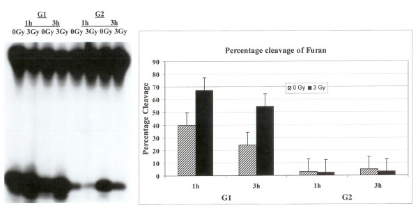
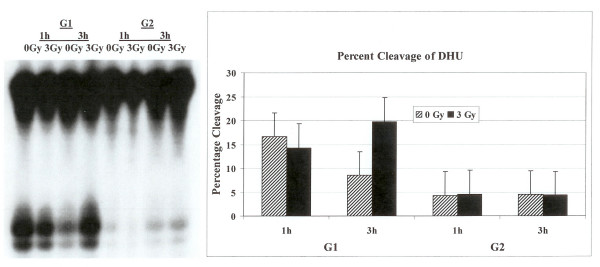
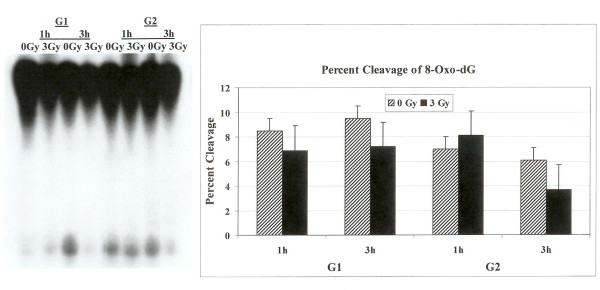
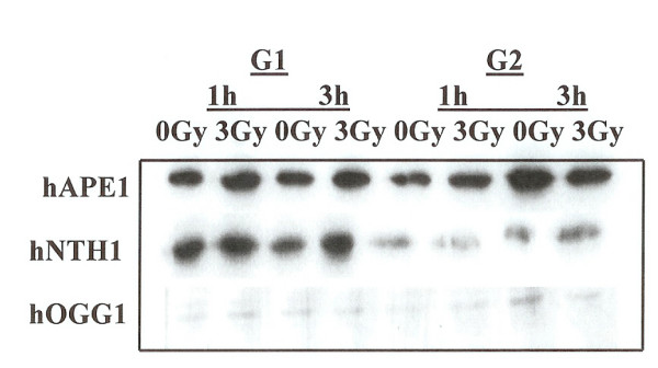
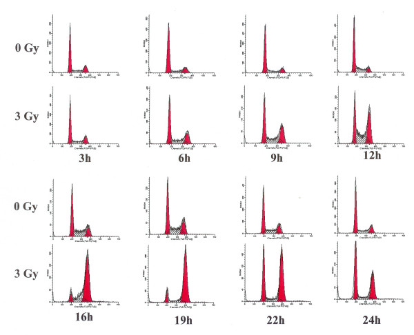
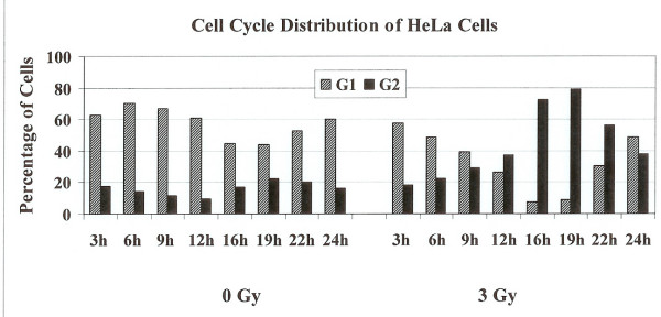
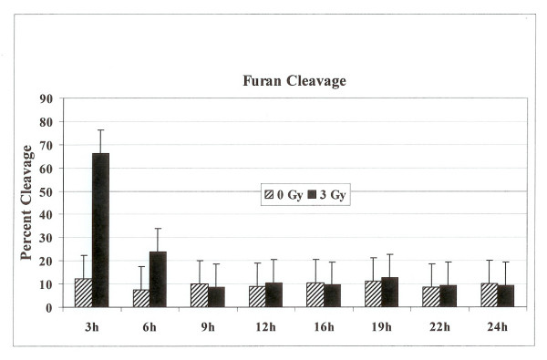
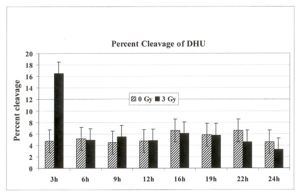
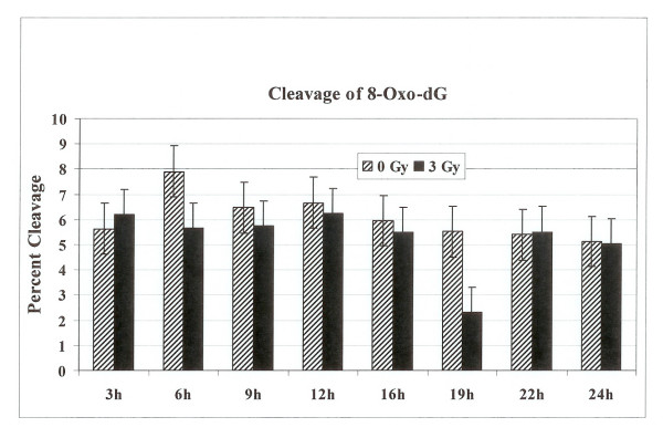
Results: Using an in vitro incision assay involving radiolabeled oligonucleotides with specific DNA lesions, we examined the activities of several BER enzymes in the whole cell extracts prepared from synchronized human HeLa cells irradiated in G1 and G2 phase of the cell cycle. The activities of human endonuclease III (hNTH1), a glycosylase/lyase that removes several damaged bases from DNA including dihydrouracil (DHU), 8-oxoguanine-DNA glycosylase (hOGG1) that recognizes 7,8-dihydro-8-oxo-2'-deoxyguanosine (8-oxoG) lesion and apurinic/apyrimidinic endonuclease (hAPE1) that acts on abasic sites including synthetic analog furan were examined.

Conclusion: Overall the repair activities of hNTH1 and hAPE1 were higher in the G1 compared to G2 phase of the cell cycle. The percent cleavages of oligonucleotide substrate with furan were greater than substrate with DHU in both G1 and G2 phases. The irradiation of cells enhanced the cleavage of substrates with furan and DHU only in G1 phase. The activity of hOGG1 was much lower and did not vary within the cell cycle. These results demonstrate the cell cycle phase dependence on the BER of ionizing radiation-induced DNA damage. Interestingly no evidence of enhanced BER activities was found in irradiated cells arrested in G2 phase.


Figures

Figure 1
Figure 1
Incision of oligonucleotide containing furan lesion. HeLa cells were synchronized and Flow cytometer analysis confirmed their G1 or G2 status. The labeled oligonucleotide was incubated with cell extracts prepared from synchronized G1 or G2 HeLa cells either irradiated or not irradiated. The incision products on the gel were quantified with PhosphorImager to determine percentage cleavage.
Figure 2
Figure 2
Cleavage of oligonucleotide containing DHU lesion. The labeled oligonucleotide was incubated with cell extracts prepared from synchronized G1 or G2 HeLa cells either not irradiated or irradiated with 3 Gy. The cleavage products on the gel were quantified with PhosphorImager.
Figure 3
Figure 3
Incision of oligonucleotide with 8-Oxo-G DNA lesion. The labeled oligonucleotide was incubated with cell extracts prepared from synchronized G1 or G2 HeLa cells either irradiated or not irradiated. The cleavage products on the gel were quantified with PhosphorImager to determine percentage cleavage.
Figure 4
Figure 4
Western blot analysis of the cells synchronized in G1 or G2. The expression of hAPE1, hNTH1 and hOGG1 was examined in G1 and G2 phase cells. Cells were irradiated with 3 Gy and the expression of these proteins was monitored at 1 hour and at 3 hours post irradiation.
Figure 5
Figure 5
Cell cycle distribution of HeLa Cells after radiation exposure. Cells were irradiated with 3 Gy and samples were taken after 3 h, 6 h, 9 h, 12 h, 16 h, 22 h, and 24 h for flow cytometry analysis. The cell cycle distribution of irradiated cells was compared to unirradiated cells collected at the same time points. The left peek in each case represents cells in G1 and the right peek represents cells in G2 phase.
Figure 6
Figure 6
Percentage of cells in G1 or G2 phases in unirradiated or 3 Gy irradiated cells collected at various time points.
Figure 7
Figure 7
The percentage cleavage of oligonucleotide substrate containing furan lesion. The radiolabeled oligonucleotide substrate was incubated with cell extracts prepared from either unirradiated or 3 Gy irradiated cells collected at various time points. The reaction products were separated on polyacrylamide gel and the incised fragments were quantified to calculate the percentage cleavage at the furan site.
Figure 8
Figure 8
The percentage cleavage of oligonucleotide substrate containing DHU lesion. The radiolabeled oligonucleotide substrate was incubated with cell extracts prepared from either unirradiated or 3 Gy irradiated cells collected at various time points. The reaction products were separated on polyacrylamide gel and the incised fragments were quantified to calculate the percentage cleavage at the DHU site.
Figure 9
Figure 9
The percentage cleavage of oligonucleotide substrate containing 8-Oxo-dG lesion. The radiolabeled oligonucleotide substrate was incubated with cell extracts prepared from either unirradiated or 3 Gy irradiated cells collected at various time points. The reaction products were separated on polyacrylamide gel and the incised fragments were quantified to calculate the percentage cleavage at the 8-Oxo-dG site.

References

    1. Swanson RL, Morey NJ, Doetsch PW, Jinks-Robertson S. Overlapping specificities of base excision repair, nucleotide excision repair, recombination, and translesion synthesis pathways for DNA base damage in Saccharomyces cerevisiae. Mol Cell Biol. 1999;19:2929–2935. - PMC - PubMed
    1. Reardon JT, Bessho T, Kung HC, Bolton PH, Sancar A. In vitro repair of oxidative DNA damage by human nucleotide excision repair system: possible explanation for neurodegeneration in xeroderma pigmentosum patients. Proc Natl Acad Sci U S A. 1997;94:9463–9468. doi: 10.1073/pnas.94.17.9463. - DOI - PMC - PubMed
    1. D'Errico M, Parlanti E, Teson M, de Jesus BM, Degan P, Calcagnile A, Jaruga P, Bjoras M, Crescenzi M, Pedrini AM, Egly JM, Zambruno G, Stefanini M, Dizdaroglu M, Dogliotti E. New functions of XPC in the protection of human skin cells from oxidative damage. Embo J. 2006;25:4305–4315. doi: 10.1038/sj.emboj.7601277. - DOI - PMC - PubMed
    1. Katafuchi A, Nakano T, Masaoka A, Terato H, Iwai S, Hanaoka F, Ide H. Differential specificity of human and Escherichia coli endonuclease III and VIII homologues for oxidative base lesions. J Biol Chem. 2004;279:14464–14471. doi: 10.1074/jbc.M400393200. - DOI - PubMed
    1. Sung JS, Demple B. Roles of base excision repair subpathways in correcting oxidized abasic sites in DNA. Febs J. 2006;273:1620–1629. doi: 10.1111/j.1742-4658.2006.05192.x. - DOI - PubMed