Comparative Study
. 2007;9(5):R97.
doi: 10.1186/ar2298.

NF-kappaB inhibitor dehydroxymethylepoxyquinomicin suppresses osteoclastogenesis and expression of NFATc1 in mouse arthritis without affecting expression of RANKL, osteoprotegerin or macrophage colony-stimulating factor

Affiliations
Comparative Study

NF-kappaB inhibitor dehydroxymethylepoxyquinomicin suppresses osteoclastogenesis and expression of NFATc1 in mouse arthritis without affecting expression of RANKL, osteoprotegerin or macrophage colony-stimulating factor

Tetsuo Kubota et al. Arthritis Res Ther. 2007.

Abstract

Inhibition of NF-kappaB is known to be effective in reducing both inflammation and bone destruction in animal models of arthritis. Our previous study demonstrated that a small cell-permeable NF-kappaB inhibitor, dehydroxymethylepoxyquinomicin (DHMEQ), suppresses expression of proinflammatory cytokines and ameliorates mouse arthritis. It remained unclear, however, whether DHMEQ directly affects osteoclast precursor cells to suppress their differentiation to mature osteoclasts in vivo. The effect of DHMEQ on human osteoclastogenesis also remained elusive. In the present study, we therefore examined the effect of DHMEQ on osteoclastogenesis using a mouse collagen-induced arthritis model, and using culture systems of fibroblast-like synovial cells obtained from patients with rheumatoid arthritis, and of osteoclast precursor cells from peripheral blood of healthy volunteers. DHMEQ significantly suppressed formation of osteoclasts in arthritic joints, and also suppressed expression of NFATc1 along the inner surfaces of bone lacunae and the eroded bone surface, while serum levels of soluble receptor activator of NF-kappaB ligand (RANKL), osteoprotegerin and macrophage colony-stimulating factor were not affected by the treatment. DHMEQ also did not suppress spontaneous expression of RANKL nor of macrophage colony-stimulating factor in culture of fibroblast-like synovial cells obtained from patients with rheumatoid arthritis. These results suggest that DHMEQ suppresses osteoclastogenesis in vivo, through downregulation of NFATc1 expression, without significantly affecting expression of upstream molecules of the RANKL/receptor activator of NF-kappaB/osteoprotegerin cascade, at least in our experimental condition. Furthermore, in the presence of RANKL and macrophage colony-stimulating factor, differentiation and activation of human osteoclasts were also suppressed by DHMEQ, suggesting the possibility of future application of NF-kappaB inhibitors to rheumatoid arthritis therapy.


Figures

Figure 1
Figure 1
Effect of dehydroxymethylepoxyquinomicin on inflammation and bone destruction in collagen-induced mouse arthritis. (a) Increase (%) of the sum of the thickness of the right and left hind paws in each mouse during day -4 and day 10. Horizontal bars represent the mean. DHMEQ, dehydroxymethylepoxyquinomicin. (b) Radiographic scores of the ankle joints were determined as described in Materials and methods, and were normalized to the normal mice. Values are expressed as the mean ± standard deviation, and represent data obtained by three independent investigators. Data were compared by Student's t test.
Figure 2
Figure 2
Effect of dehydroxymethylepoxyquinomicin on differentiation of osteoclasts in ankle joints of mice with collagen-induced arthritis. After taking radiographs (a-c), the ankle joints were histochemically examined for tartrate-resistant acid phosphatase-positive cells (d-i). (a), (d) and (g) Typical joint of an arthritic mouse treated with vehicle alone. (b), (e) and (h) Typical joint of an arthritic mouse treated with dehydroxymethylepoxyquinomicin. (c), (f) and (i) Joint of an age-matched normal mouse. Arrow, multinucleated giant osteoclasts.
Figure 3
Figure 3
Quantitative estimation of the suppressive effect of dehydroxymethylepoxyquinomicin on in vivo osteoclastogenesis. The mean number of tartrate-resistant acid phosphatase-positive giant cells with four or more nuclei in the individual ankle joints of arthritic mice treated with vehicle alone (n = 13), of mice treated with dehydroxymethylepoxyquinomicin (DHMEQ) (n = 12), and of normal mice (n = 6) were counted under a microscope by two investigators in a blinded manner to the assignment of mouse groups. The results shown are the mean ± standard error of the mean of four independent counts, and were compared by Student's t test.
Figure 4
Figure 4
Effect of dehydroxymethylepoxyquinomicin on serum factors involved in osteoclastogenesis. Effect of dehydroxymethylepoxyquinomicin (DHMEQ) on serum levels of (a) osteoprotegerin (OPG), (b) soluble receptor activator of NF-κB ligand (sRANKL), (c) sRANKL/OPG ratio and (d) macrophage colony-stimulating factor. Serum levels of these cytokines in individual arthritic mice 3 hours after the last treatment with vehicle alone (n = 13) or with DHMEQ (n = 12), and in age-matched normal mice (n = 4–6), were determined by ELISA. Horizontal lines represent the median. Data were analyzed by the Mann-Whitney test. P < 0.05 was considered significant; ns, not significant.
Figure 5
Figure 5
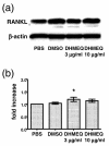
Effect of dehydroxymethylepoxyquinomicin on human fibroblast-like synovial cells. Effect of dehydroxymethylepoxyquinomicin (DHMEQ) on expression of receptor activator of NF-κB ligand (RANKL) and of macrophage colony-stimulating factor (M-CSF) by fibroblast-like synovial cells obtained from patients with rheumatoid arthritis (RA-FLS). The RA-FLS were incubated with DHMEQ, with vehicle (dimethyl sulfoxide (DMSO)), or with PBS for 24 hours. (a) Cell lysates were analyzed by western blotting with anti-RANKL or with anti-β-actin monoclonal antibody. Representative data of similar results obtained using cell lines from two patients with RA are shown. (b) Concentration of M-CSF in the culture supernatant measured by ELISA; results expressed as relative values compared with PBS. Data are the mean ± standard error of the mean of independent experiments carried out in triplicate using cell lines obtained from six patients with RA, and were compared by Student's t test. *P < 0.05.
Figure 6
Figure 6
Effect of dehydroxymethylepoxyquinomicin on NF-κB activation and NFATc1 expression in joints of collagen-induced arthritis. Fresh frozen sections of each ankle joint were double-stained: (a, d, g) with FITC-labeled antibody to an activated form of the p65 subunit of NF-κB, and (b, e, h) with phycoerythrin-labeled antibody to NFATc1. (c, f, i) Transmission microscopy images of the same slides to show the articular structure. First row, a typical joint of an arthritic mouse treated with vehicle alone (a-c). Second row, a typical joint of an arthritic mouse treated with dehydroxymethylepoxyquinomicin (d-f). Third row, a joint of an age-matched normal mouse (g-i). White arrow (a), staining by anti-NF-κB p65 antibody; blue arrow (b), staining by anti-NFATc1 antibody of the cells along the inner surfaces of bone lacunae.
Figure 7
Figure 7
Effect of dehydroxymethylepoxyquinomicin on human osteoclastogenesis and production of matrix metalloprotease-9 by human osteoclasts. (a) Peripheral blood monocytes were incubated in 96-well plates with macrophage colony-stimulating factor (M-CSF), receptor activator of NF-κB ligand (RANKL), and the indicated concentrations of dehydroxymethylepoxyquinomicin (DHMEQ). At day 7, the total number of tartrate-resistant acid phosphatase (TRAP)-positive multinucleated cells (MNC) with three or more nuclei/well was counted. Representative data of three independent experiments are shown. *P < 0.01, DHMEQ versus dimethyl sulfoxide (DMSO). (b) Peripheral blood monocytes were incubated in 96-well plates with M-CSF and RANKL without DHMEQ. At day 7, the medium was replaced with fresh medium and the indicated concentrations of DHMEQ were added. The culture supernatant was collected at day 8, and the matrix metalloprotease-9 (MMP-9) concentration was measured by ELISA. Representative data of two independent experiments are shown. Data represent the mean ± standard error of the mean of triplicate wells, and were compared by Student's t test. *P < 0.05, DHMEQ versus DMSO.

References

    1. Lipsky PE, van der Heijde DM, St Clair EW, Furst DE, Breedveld FC, Kalden JR, Smolen JS, Weisman M, Emery P, Feldmann M, et al. Infliximab and methotrexate in the treatment of rheumatoid arthritis. Anti-tumor necrosis factor trial in rheumatoid arthritis with concomitant therapy study group. N Engl J Med. 2000;343:1594–1602. doi: 10.1056/NEJM200011303432202. - DOI - PubMed
    1. van der Heijde D, Klareskog L, Rodriguez-Valverde V, Codreanu C, Bolosiu H, Melo-Gomes J, Tornero-Molina J, Wajdula J, Pedersen R, Fatenejad S. Comparison of etanercept and methotrexate, alone and combined, in the treatment of rheumatoid arthritis. Two-year clinical and radiographic results from the TEMPO study, a double-blind, randomized trial. Arthritis Rheum. 2006;54:1063–1074. doi: 10.1002/art.21655. - DOI - PubMed
    1. Smolen JS, van der Heijde DMFM, St Clair EW, Emery P, Bathon JM, Keystone E, Maini RN, Kalden JR, Schiff M, Baker D, et al. Predictors of joint damage in patients with early rheumatoid arthritis treated with high-dose methotrexate with or without concomitant infliximab. Results from the ASPIRE trial. Arthritis Rheum. 2006;54:702–710. doi: 10.1002/art.21678. - DOI - PubMed
    1. Redlich K, Hayer S, Ricci R, David J-P, Tohidast-Akrad M, Kollias G, Steiner G, Smolen JS, Wagner EF, Schett G. Osteoclasts are essential for TNF-α-mediated joint destruction. J Clin Invest. 2002;110:1419–1427. doi: 10.1172/JCI200215582. - DOI - PMC - PubMed
    1. Teitelbaum SL. Bone resorption by osteoclasts. Science. 2000;289:1504–1508. doi: 10.1126/science.289.5484.1504. - DOI - PubMed

Publication types

MeSH terms

LinkOut - more resources