. 2007 Oct 1;204(10):2407-22.
doi: 10.1084/jem.20070628. Epub 2007 Sep 24.

Selective predisposition to bacterial infections in IRAK-4-deficient children: IRAK-4-dependent TLRs are otherwise redundant in protective immunity

Affiliations

Selective predisposition to bacterial infections in IRAK-4-deficient children: IRAK-4-dependent TLRs are otherwise redundant in protective immunity

Cheng-Lung Ku et al. J Exp Med. .

Abstract

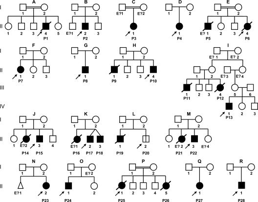
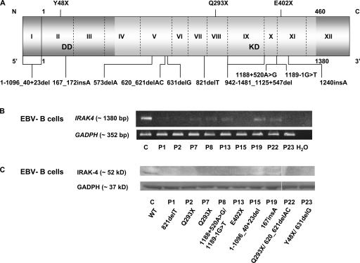
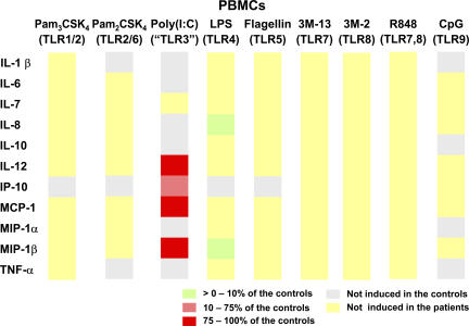
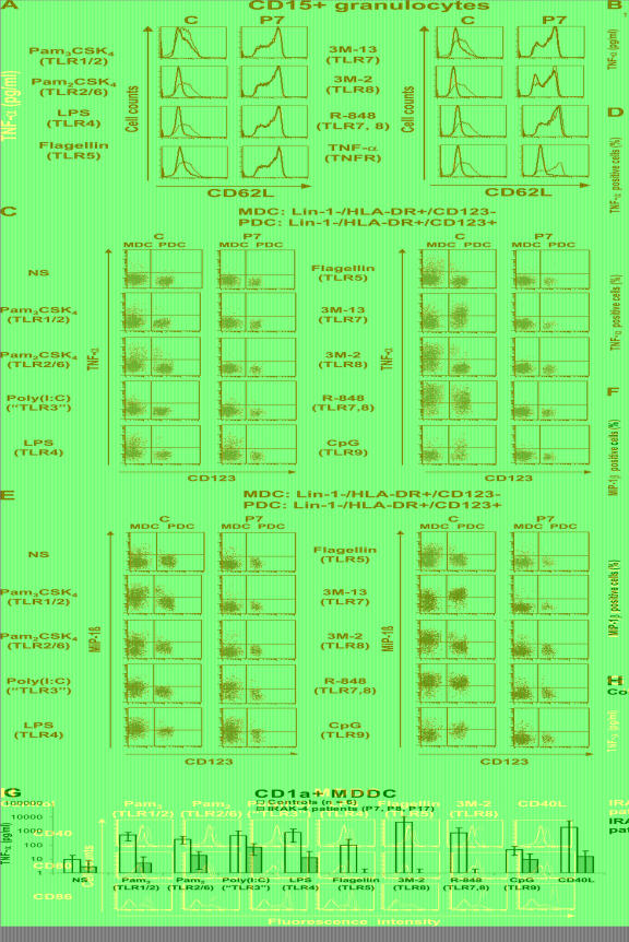
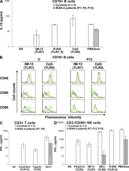
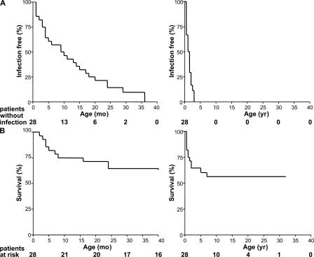
Human interleukin (IL) 1 receptor-associated kinase 4 (IRAK-4) deficiency is a recently discovered primary immunodeficiency that impairs Toll/IL-1R immunity, except for the Toll-like receptor (TLR) 3- and TLR4-interferon (IFN)-alpha/beta pathways. The clinical and immunological phenotype remains largely unknown. We diagnosed up to 28 patients with IRAK-4 deficiency, tested blood TLR responses for individual leukocyte subsets, and TLR responses for multiple cytokines. The patients' peripheral blood mononuclear cells (PBMCs) did not induce the 11 non-IFN cytokines tested upon activation with TLR agonists other than the nonspecific TLR3 agonist poly(I:C). The patients' individual cell subsets from both myeloid (granulocytes, monocytes, monocyte-derived dendritic cells [MDDCs], myeloid DCs [MDCs], and plasmacytoid DCs) and lymphoid (B, T, and NK cells) lineages did not respond to the TLR agonists that stimulated control cells, with the exception of residual responses to poly(I:C) and lipopolysaccharide in MDCs and MDDCs. Most patients (22 out of 28; 79%) suffered from invasive pneumococcal disease, which was often recurrent (13 out of 22; 59%). Other infections were rare, with the exception of severe staphylococcal disease (9 out of 28; 32%). Almost half of the patients died (12 out of 28; 43%). No death and no invasive infection occurred in patients older than 8 and 14 yr, respectively. The IRAK-4-dependent TLRs and IL-1Rs are therefore vital for childhood immunity to pyogenic bacteria, particularly Streptococcus pneumoniae. Conversely, IRAK-4-dependent human TLRs appear to play a redundant role in protective immunity to most infections, at most limited to childhood immunity to some pyogenic bacteria.


Figures

Figure 1.
Figure 1.
Pedigree of the 18 kindreds identified with IRAK-4 deficiency. Each kindred is designated by a capital letter (A–R), each generation is designated by a Roman numeral (I–IV), and each individual is designated by an Arabic numeral (from left to right). IRAK-4–deficient patients with a clinical phenotype are represented as closed symbols. P20, the only patient with confirmed IRAK-4 deficiency but no known clinical phenotype, is represented with an open square divided by a black line. In each family, the proband is indicated by an arrow. Individuals whose genetic status could not be evaluated are indicated by “E?”; they include four individuals (P5, 14, 16, and 21) thought to be IRAK-4 deficient based on their clinical phenotypes.
Figure 2.
Figure 2.
IRAK-4 deficiency. (A) Schematic representation of IRAK4 with all identified mutations. The gene is composed of 12 exons, with exon 1 and a part of exon 12 noncoding. The N-terminal death domain (DD) and C-terminal kinase domain (KD) are shown in light gray. (B) RT-PCR of the full-length IRAK4 and GAPDH genes in B-EBVs from a healthy control (C) and nine IRAK-4–deficient patients. (C) IRAK-4 and GAPDH protein levels in B-EBVs from a healthy control and nine IRAK-4–deficient patients, as shown by Western blotting. White lines indicate that intervening lanes have been spliced out.
Figure 3.
Figure 3.
Impaired cellular responses to TIR agonists in IRAK-4–deficient cell lines. (A) TNF-α production by B-EBVs from a healthy control (C) and seven IRAK-4–deficient patients 24 h after stimulation with various TLR agonists and PMA/ionomycin. (B) IL-6 production by SV40-fibroblasts from a healthy control and eight IRAK-4–deficient patients after 24 h of stimulation with IL-1β, TNF-α, poly(I:C), and PMA/ionomycin. Mean values and SDs are shown for triplicates of a single experiment.
Figure 4.
Figure 4.
Multiple cytokine secretion in IRAK-4–deficient PBMCs. PBMCs from three healthy controls and three IRAK-4–deficient patients (P17, 18, and 22) were activated with various TLR agonists for 24 h. Cytokine levels are represented as ratios of the mean secretion observed in the three IRAK-4–deficient patients to that in three healthy controls. Cytokines represented in gray are not induced upon the stimulation of control PBMCs.
Figure 5.
Figure 5.
Impaired responses to TLR agonists in IRAK-4–deficient individual myeloid subsets. (A) Cleavage of CD62 ligand (CD62L) at the surface of granulocytes from a healthy control and an IRAK-4–deficient patient (P7) after activation for 1 h with various TLR agonists and TNF-α. The black line shows CD62L expression on nonactivated granulocytes, and the red line shows CD62L expression after 1 h of activation with various agonists (induced CD62L shedding). One experiment representative of four (P7, 8, 13, and 15) is shown. (B) TNF-α secretion by CD14+ monocytes after 24 h of activation with various TLR agonists. Mean values and SDs were calculated from four healthy controls and three IRAK-4–deficient patients. (C–F) Ex vivo MDC and PDC responses. PBMCs from healthy controls and IRAK-4–deficient patients were stimulated with various TLR agonists. In both subsets, responses were measured by staining for intracellular TNF-α (C) and MIP-1β (E). Mean values and SDs were calculated from six different controls and four IRAK-4–deficient patients for TNF-α (D), and from seven different controls and three IRAK-4–deficient patients for MIP-1β (F). (G) TNF-α secretion in vitro by MDDCs after 24 h of activation. Means and SDs were calculated from six different controls and three different IRAK-4–deficient patients. (H) Induction of CD40, CD80, and CD86 surface expression on MDDCs from a control (top) and an IRAK-4–deficient patient (bottom) after 24 h of stimulation with various TLR agonists. Black and green lines indicate the expression of CD40, CD80, and CD86 without and after stimulation, respectively. The experiment shown is representative of three independent experiments (also performed on patients P15 and 18). C, control.
Figure 6.
Figure 6.
Lack of response to TLR agonists of individual IRAK-4–deficient lymphoid subsets. (A) IL-10 secretion by CD19+ B cells after 24 h of activation with various TLR agonists and PMA/ionomycin. Mean values ± SD were calculated from the data obtained for three different controls and three IRAK-4–deficient patients. (B) Induction of CD40, CD80, and CD86 surface expression on CD19+ B cells after activation for 72 h with 3M-13 and CpG. Black and green lines indicate the expression of CD40, CD80, and CD86 without and after stimulation, respectively. Data are representative of two independent experiments. (C) IFN-γ secretion by CD3+ T cells after stimulation for 24 h with various TLR agonists and anti-CD3 (50 ng/ml OKT3) antibody in the presence of 100 U/ml IL-2 for 2 d. Mean values ± SD were calculated for three different controls and two IRAK-4–deficient patients. (D) IFN-γ secretion by CD3/CD56+ NK cells after activation for 24 h with various TLR agonists and PMA/ionomycin. Mean values and SDs were calculated for three different controls and three IRAK-4–deficient patients.
Figure 7.
Figure 7.
Epidemiological features of IRAK-4 deficiency. (A) Incidence of invasive infections in IRAK-4–deficient patients during the first 40 mo of life (left) and the first 40 yr of life (right). Invasive infections included meningitis, septicemia, and arthritis. (B) Survival curve of 28 IRAK-4–deficient patients during the first 40 mo of life (left) and the first 40 yr of life (right).

References

    1. Picard, C., A. Puel, M. Bonnet, C.L. Ku, J. Bustamante, K. Yang, C. Soudais, S. Dupuis, J. Feinberg, C. Fieschi, et al. 2003. Pyogenic bacterial infections in humans with IRAK-4 deficiency. Science. 299:2076–2079. - PubMed
    1. Kuhns, D.B., D.A. Long Priel, and J.I. Gallin. 1997. Endotoxin and IL-1 hyporesponsiveness in a patient with recurrent bacterial infections. J. Immunol. 158:3959–3964. - PubMed
    1. Haraguchi, S., N.K. Day, R.P. Nelson Jr., P. Emmanuel, J.E. Duplantier, C.S. Christodoulou, and R.A. Good. 1998. Interleukin 12 deficiency associated with recurrent infections. Proc. Natl. Acad. Sci. USA. 95:13125–13129. - PMC - PubMed
    1. Medvedev, A.E., A. Lentschat, D.B. Kuhns, J.C. Blanco, C. Salkowski, S. Zhang, M. Arditi, J.I. Gallin, and S.N. Vogel. 2003. Distinct mutations in IRAK-4 confer hyporesponsiveness to lipopolysaccharide and interleukin 1 in a patient with recurrent bacterial infections. J. Exp. Med. 198:521–531. - PMC - PubMed
    1. Currie, A.J., D.J. Davidson, G.S. Reid, S. Bharya, K.L. MacDonald, R.S. Devon, and D.P. Speert. 2004. Primary immunodeficiency to pneumococcal infection due to a defect in Toll-like receptor signaling. J. Pediatr. 144:512–518. - PubMed

Publication types

MeSH terms