. 2007 Oct 1;204(10):2461-71.
doi: 10.1084/jem.20070818. Epub 2007 Sep 24.

Staphylococcal complement evasion by various convertase-blocking molecules

Affiliations

Staphylococcal complement evasion by various convertase-blocking molecules

Ilse Jongerius et al. J Exp Med. .

Abstract

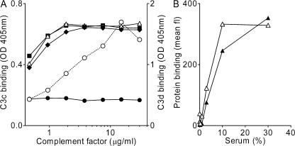
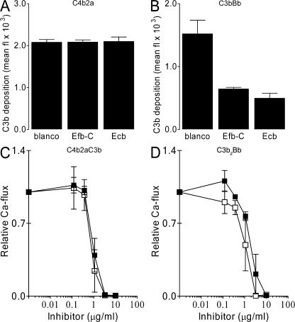
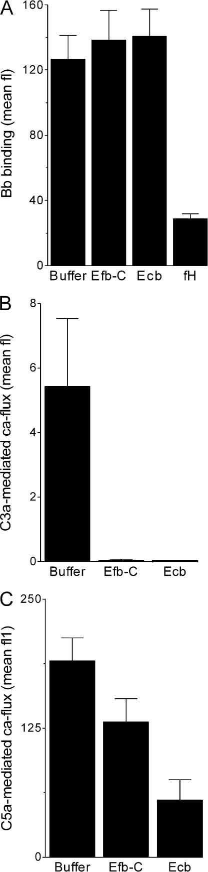
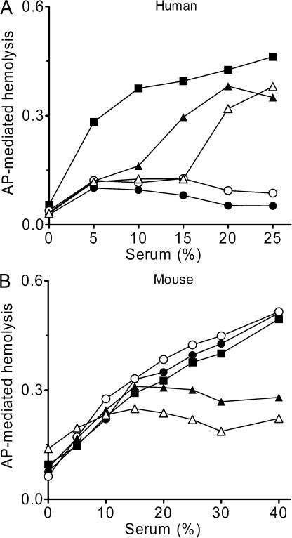
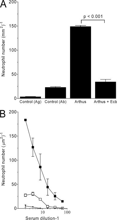
To combat the human immune response, bacteria should be able to divert the effectiveness of the complement system. We identify four potent complement inhibitors in Staphylococcus aureus that are part of a new immune evasion cluster. Two are homologues of the C3 convertase modulator staphylococcal complement inhibitor (SCIN) and function in a similar way as SCIN. Extracellular fibrinogen-binding protein (Efb) and its homologue extracellular complement-binding protein (Ecb) are identified as potent complement evasion molecules, and their inhibitory mechanism was pinpointed to blocking C3b-containing convertases: the alternative pathway C3 convertase C3bBb and the C5 convertases C4b2aC3b and C3b2Bb. The potency of Efb and Ecb to block C5 convertase activity was demonstrated by their ability to block C5a generation and C5a-mediated neutrophil activation in vitro. Further, Ecb blocks C5a-dependent neutrophil recruitment into the peritoneal cavity in a mouse model of immune complex peritonitis. The strong antiinflammatory properties of these novel S. aureus-derived convertase inhibitors make these compounds interesting drug candidates for complement-mediated diseases.


Figures

Figure 1.
Figure 1.
A new IEC in S. aureus. Graphic representation of the novel IEC-2 in sequenced S. aureus strains. Black arrows indicate known or putative immune evasion molecules: Ecb (ecb), FLIPr (flr), FLIPr-like (fll), Efb (efb), SCIN-B (scb), SCIN-C (scc), α-hemolysin (hla), SSL12 (ssl12), SSL13 (ssl13), and SSL14 (ssl14). Genes with unknown functions are named according to their locus number in S. aureus N315. All strains carry either flr (black) or fll (dark gray) and scb (black) or scc (dark gray). The household genes (light gray) murI (glutamate racemase), SA998, SA999, and argF (ornithine carbamoyltransferase) form the borders of IEC-2. White arrows delineate ORFs similar to bacteriophage proteins. Three transposases for insertion sequences were found: SA1006 in N315 and Mu50, and SAR1138 in MRSA252. S. aureus strain RF122 represents a bovine isolate.
Figure 2.
Figure 2.
Innate immune evasion by four putative complement inhibitors on IEC-2. (A) SCIN-B and SCIN-C inhibit C5a production. S. aureus was incubated with 10% human serum in the presence of 10 μg/ml SCIN (□), SCIN-B (•), SCIN-C (○), or ORF-D (▪). C5a formation was measured by using supernatants as stimuli for calcium mobilization of human neutrophils. SCIN-B and SCIN-C show a dose-dependent inhibition of C5a formation, whereas ORF-D had no effect. (B) Efb-C and Ecb block C5a production during opsonization. Dose-dependent inhibition of C5a-mediated neutrophil activation by Efb-C (▴), Ecb (Δ), and SCIN (□). (C) Supernatants that were activated in the presence of 10 μg/ml SCIN, SCIN-B, SCIN-C, Efb-C, or Ecb were less potent in up-regulating CD11b expression on neutrophils. (D) Supernatants that were activated in the presence of 10 μg/ml SCIN, SCIN-B, SCIN-C, Efb-C, or Ecb were less potent in down-regulation of CD62L expression on neutrophils. (E) SCIN (□), SCIN-B (•), and SCIN-C (○) inhibit phagocytosis of S. aureus by human neutrophils in 10% human serum. ORF-D (▪), Efb-C (▴), and Ecb (Δ) did not affect phagocytosis. (F) SCIN, SCIN-B, and SCIN-C inhibit C3b/iC3b deposition on the bacterial surface in 10% human serum, whereas Efb-C and Ecb do not influence opsonization. (G) Antibody titers against SCIN, SCIN-B, SCIN-C, Efb, and Ecb in sera of 12 healthy lab volunteers. Horizontal lines represent the mean. All data represent the mean ± SE of three separate experiments. Mean fl, mean fluorescence.
Figure 3.
Figure 3.
SCIN-B and SCIN-C function similar to SCIN. ELISA experiments showing that SCIN (□), SCIN-B (•), and SCIN-C (○) inhibit C3b deposition after CP (5% serum; A), LP (5% serum; B), and AP activation (30% serum; C). SCIN, SCIN-B, and SCIN-C also prevent C5b-9 deposition during CP (5% serum; D), LP (5% serum; E), and AP activation (30% serum; F). ORF-D (▪) cannot inhibit C3b or C5b-9 deposition in ELISA. Data shown in A–F represent the mean ± SE of three separate experiments. (G and H) SCIN, SCIN-B, and SCIN-C enhance convertase stability during opsonization of S. aureus in 20% human serum. Opsonization was performed for 0, 2, 10, and 30 min. Surface-bound convertases were detected by immunoblotting using anti-C2 (G) or anti-fB (H) antibodies. Blots represent three separate experiments.
Figure 4.
Figure 4.
Efb-C and Ecb act differently than SCIN. ELISA experiments showing that Efb-C (▴) and Ecb (Δ) do not inhibit C3b deposition after CP (5% serum; A) or LP (5% serum; B) activation but strongly block C3b deposition after AP activation (30% serum; C). Efb-C and Ecb strongly prevent C5b-9 deposition in response to activation of all pathways: CP (5% serum; D), LP (5% serum; E), and AP (30% serum; F). Data shown in A–F are the mean ± SE of three separate experiments. (G and H) Efb-C and Ecb do not enhance convertase stability during opsonization of S. aureus in 20% human serum. Opsonization was performed for 0, 2, 10, and 30 min. Surface-bound convertases were detected by immunoblotting using anti-C2 (G) or anti-fB (H) antibodies. Blots represent three separate experiments.
Figure 5.
Figure 5.
Complement-binding properties of Efb-C and Ecb. (A) Ecb binds the C3d domain of C3 (fragments). 5 μg/ml Ecb was coated to ELISA plates, and subsequent binding of C3 (▪), iC3b (Δ), C3b (♦), C3c (•), and C3d (○) was determined. Ecb exclusively binds C3d-containing C3 molecules. (B) Efb-C (▴) and Ecb (Δ) bind to S. aureus in a serum-dependent manner. 10 μg/ml of His-tagged Efb-C and Ecb were incubated with S. aureus and human serum for 30 min at 37°C, and binding was detected using anti-His antibodies and flow cytometry. His-tagged SSL7 served as a negative control. Graphs show one representative figure out of three separate experiments. Mean fl, mean fluorescence.
Figure 6.
Figure 6.
Efb-C and Ecb act on C3b-containing convertases. Efb-C and Ecb specifically block C3b-containing convertases. S. aureus was incubated with 10% fD-depleted serum (to measure CP and LP activation) or 10% human serum in the presence of Mg-EGTA (to measure AP activation). Complement activation was measured at the level of C3b deposition by flow cytometry using anti-C3 antibodies (A and B) or at the level of C5a production by calcium mobilization (C and D). Efb-C (▪) and Ecb (□) do not prevent C3b deposition by the CP/LP (A) but inhibit C3b deposition by the AP (B). Formation of C5a was blocked in response to CP and LP activation (C), as well as AP activation (D). Data shown represent the mean ± SE of three separate experiments. Mean fl, mean fluorescence.
Figure 7.
Figure 7.
Efb-C and Ecb inhibit C3 and C5 cleavage by convertases. (A) Efb-C and Ecb do not promote decay of convertases. AP convertases were created on the bacterial surface using purified components. Subsequent incubation with inhibitors showed that Efb-C and Ecb do not dissociate convertases like fH. Surface-bound convertases were detected using anti-Bb antibodies and flow cytometry. (B and C) Efb-C and Ecb inhibit convertase activity. Zymosan-bound convertases were incubated with purified C3 or C5, and generated C3a or C5a was measured in a neutrophil calcium mobilization assay. (B) Efb-C and Ecb inhibit C3 cleavage by AP convertases. (C) Efb-C and Ecb inhibit C5 cleavage by convertases. Data shown represent the mean ± SE of three separate experiments. Mean fl, mean fluorescence.
Figure 8.
Figure 8.
Efb-C and Ecb are not human specific. (A) SCIN-B, SCIN-C, Efb-C, and Ecb inhibit AP-mediated hemolysis of rabbit red blood cells in human serum. SCIN-B (○) and SCIN-C (•) completely block hemolysis at high serum concentrations. Efb-C (▴) and Ecb (Δ) also inhibit AP-mediated hemolysis but are less effective than SCIN-B or SCIN-C (all inhibitors at 10μg/ml). (B) Efb-C and Ecb inhibit AP-mediated hemolysis in mouse serum. Addition of 10 μg/ml Efb-C (▴) or Ecb (Δ) results in strong inhibition of MAC formation in mouse serum, whereas SCIN-B (○) and SCIN-C (•) had no effect. Graphs show one representative figure out of three separate experiments.
Figure 9.
Figure 9.
Ecb completely blocks IC-induced neutrophil influx in vivo. (A) In vivo complement inhibition by Ecb was tested in the reverse passive Arthus reaction peritonitis model. Neutrophils accumulate in the peritoneal cavity within 6 h after IC challenge. i.p. and i.v. injection of Ecb before IC challenge resulted in complete inhibition of neutrophil migration. Injection of Ecb alone did not induce IC peritonitis. P < 0.001 by analysis of variance (n = 10 mice per group). (B) Ecb completely blocks C5a-dependent migration of BM-derived mouse neutrophils in vitro. Preincubation of zymosan with C5-sufficient (▪) but not C5-deficient plasma (▾) results in neutrophil migration. In the presence of 50 μg/ml Ecb, C5a production in C5-sufficient plasma was blocked (□). Data shown represent the mean ± SE of three separate experiments.

Similar articles

Cited by

References

    1. Neth, O., D.L. Jack, M. Johnson, N.J. Klein, and M.W. Turner. 2002. Enhancement of complement activation and opsonophagocytosis by complexes of mannose-binding lectin with mannose-binding lectin-associated serine protease after binding to Staphylococcus aureus. J. Immunol. 169:4430–4436. - PubMed
    1. Wergeland, H., C. Endresen, O.B. Natas, P. Aasjord, and P. Oeding. 1984. Antibodies to Staphylococcus aureus peptidoglycan and lipoteichoic acid in sera from blood donors and patients with staphylococcal infections. Acta Pathol. Microbiol. Immunol. Scand. [B]. 92:265–269. - PubMed
    1. Walport, M.J. 2001. Complement. Second of two parts. N. Engl. J. Med. 344:1140–1144. - PubMed
    1. Walport, M.J. 2001. Complement. First of two parts. N. Engl. J. Med. 344:1058–1066. - PubMed
    1. Muller-Eberhard, H.J. 1986. The membrane attack complex of complement. Annu. Rev. Immunol. 4:503–528. - PubMed

Publication types

MeSH terms

Substances

Associated data