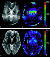
Quantitative assessment of iron accumulation in the deep gray matter of multiple sclerosis by magnetic field correlation imaging
- PMID: 17893225
- PMCID: PMC8134218
- DOI: 10.3174/ajnr.A0646
Quantitative assessment of iron accumulation in the deep gray matter of multiple sclerosis by magnetic field correlation imaging
Abstract
Background and purpose: Deposition of iron has been recognized recently as an important factor of pathophysiologic change including neurodegenerative processes in multiple sclerosis (MS). We propose that there is an excess accumulation of iron in the deep gray matter in patients with MS that can be measured with a newly developed quantitative MR technique--magnetic field correlation (MFC) imaging.
Materials and methods: With a 3T MR system, we studied 17 patients with relapsing-remitting MS and 14 age-matched healthy control subjects. We acquired MFC imaging using an asymmetric single-shot echo-planar imaging sequence. Regions of interest were selected in both deep gray matter and white matter regions, and the mean MFC values were compared between patients and controls. We also correlated the MFC data with lesion load and neuropsychologic tests in the patients.
Results: MFC measured in the deep gray matter in patients with MS was significantly higher than that in the healthy controls (P < or = .03), with an average increase of 24% in the globus pallidus, 39.5% in the putamen, and 30.6% in the thalamus. The increased iron deposition measured with MFC in the deep gray matter in the patients correlated positively with the total number of MS lesions (thalamus: r = 0.61, P = .01; globus pallidus: r = 0.52, P = .02). A moderate but significant correlation between the MFC value in the deep gray matter and the neuropsychologic tests was also found.
Conclusion: Quantitative measurements of iron content with MFC demonstrate increased accumulation of iron in the deep gray matter in patients with MS, which may be associated with the disrupted iron outflow pathway by lesions. Such abnormal accumulation of iron may contribute to neuropsychologic impairment and have implications for neurodegenerative processes in MS.
Figures


Similar articles
-
Relationship between iron accumulation and white matter injury in multiple sclerosis: a case-control study.J Neurol. 2015 Feb;262(2):402-9. doi: 10.1007/s00415-014-7569-3. Epub 2014 Nov 22. J Neurol. 2015. PMID: 25416468 Free PMC article.
-
Cognitive Implications of Deep Gray Matter Iron in Multiple Sclerosis.AJNR Am J Neuroradiol. 2017 May;38(5):942-948. doi: 10.3174/ajnr.A5109. Epub 2017 Feb 23. AJNR Am J Neuroradiol. 2017. PMID: 28232497 Free PMC article.
-
Brain iron quantification in mild traumatic brain injury: a magnetic field correlation study.AJNR Am J Neuroradiol. 2011 Nov-Dec;32(10):1851-6. doi: 10.3174/ajnr.A2637. Epub 2011 Sep 1. AJNR Am J Neuroradiol. 2011. PMID: 21885717 Free PMC article.
-
Iron Mapping in Multiple Sclerosis.Neuroimaging Clin N Am. 2017 May;27(2):335-342. doi: 10.1016/j.nic.2016.12.003. Epub 2017 Jan 20. Neuroimaging Clin N Am. 2017. PMID: 28391790 Review.
-
Multispectral quantitative magnetic resonance imaging of brain iron stores: a theoretical perspective.Top Magn Reson Imaging. 2006 Feb;17(1):19-30. doi: 10.1097/01.rmr.0000245460.82782.69. Top Magn Reson Imaging. 2006. PMID: 17179894 Review.
Cited by
-
Fluid-attenuated inversion recovery hypointensity of the pulvinar nucleus of patients with Alzheimer disease: its possible association with iron accumulation as evidenced by the t2(*) map.Korean J Radiol. 2012 Nov-Dec;13(6):674-83. doi: 10.3348/kjr.2012.13.6.674. Epub 2012 Oct 12. Korean J Radiol. 2012. PMID: 23118565 Free PMC article.
-
Iron-Insensitive Quantitative Assessment of Subcortical Gray Matter Demyelination in Multiple Sclerosis Using the Macromolecular Proton Fraction.AJNR Am J Neuroradiol. 2018 Apr;39(4):618-625. doi: 10.3174/ajnr.A5542. Epub 2018 Feb 8. AJNR Am J Neuroradiol. 2018. PMID: 29439122 Free PMC article.
-
Multimodal MR imaging of brain iron in attention deficit hyperactivity disorder: a noninvasive biomarker that responds to psychostimulant treatment?Radiology. 2014 Aug;272(2):524-32. doi: 10.1148/radiol.14140047. Epub 2014 Jun 17. Radiology. 2014. PMID: 24937545 Free PMC article.
-
Mapping of thalamic magnetic susceptibility in multiple sclerosis indicates decreasing iron with disease duration: A proposed mechanistic relationship between inflammation and oligodendrocyte vitality.Neuroimage. 2018 Feb 15;167:438-452. doi: 10.1016/j.neuroimage.2017.10.063. Epub 2017 Oct 31. Neuroimage. 2018. PMID: 29097315 Free PMC article.
-
Integrated elemental analysis supports targeting copper perturbations as a therapeutic strategy in multiple sclerosis.Neurotherapeutics. 2024 Sep;21(5):e00432. doi: 10.1016/j.neurot.2024.e00432. Epub 2024 Aug 19. Neurotherapeutics. 2024. PMID: 39164165 Free PMC article.
References
-
- Minagar A. Gray matter involvement in multiple sclerosis: a new window into pathogenesis. J Neuroimaging 2003;13:291–92 - PubMed
-
- Filippi M. Multiple sclerosis: a white matter disease with associated gray matter damage. J Neurol Sci 2001;185:3–4 - PubMed
-
- Cifelli A, Arridge M, Jezzard P, et al. Thalamic neurodegeneration in multiple sclerosis. Ann Neurol 2002;52:650–53 - PubMed
-
- Wylezinska M, Cifelli A, Jezzard P, et al. Thalamic neurodegeneration in relapsing-remitting multiple sclerosis. Neurology 2003;60:1949–54 - PubMed
-
- Inglese M, Liu S, Babb JS, et al. Three-dimensional proton spectroscopy of deep gray matter nuclei in relapsing-remitting MS. Neurology 2004;631:170–72 - PubMed
Publication types
MeSH terms
Substances
Grants and funding
LinkOut - more resources
Full Text Sources
Other Literature Sources
Medical
