. 2007 Sep 24;178(7):1265-78.
doi: 10.1083/jcb.200705021.

The type III effector EspF coordinates membrane trafficking by the spatiotemporal activation of two eukaryotic signaling pathways

Affiliations

The type III effector EspF coordinates membrane trafficking by the spatiotemporal activation of two eukaryotic signaling pathways

Neal M Alto et al. J Cell Biol. .

Abstract

Bacterial toxins and effector proteins hijack eukaryotic enzymes that are spatially localized and display rapid signaling kinetics. However, the molecular mechanisms by which virulence factors engage highly dynamic substrates in the host cell environment are poorly understood. Here, we demonstrate that the enteropathogenic Escherichia coli (EPEC) type III effector protein EspF nucleates a multiprotein signaling complex composed of eukaryotic sorting nexin 9 (SNX9) and neuronal Wiskott-Aldrich syndrome protein (N-WASP). We demonstrate that a specific and high affinity association between EspF and SNX9 induces membrane remodeling in host cells. These membrane-remodeling events are directly coupled to N-WASP/Arp2/3-mediated actin nucleation. In addition to providing a biochemical mechanism of EspF function, we find that EspF dynamically localizes to membrane-trafficking organelles in a spatiotemporal pattern that correlates with SNX9 and N-WASP activity in living cells. Thus, our findings suggest that the EspF-dependent assembly of SNX9 and N-WASP represents a novel form of signaling mimicry used to promote EPEC pathogenesis and gastrointestinal disease.


Figures

Figure 1.
Figure 1.
The EPEC effector EspF binds both SNX9 and N-WASP. (A) Schematic of EspF proline-rich repeat (PRR) domains from EPEC, EHEC 0157:H7, and Citrobacter rodentium. SNX9 (blue) and putative N-WASP (orange) binding sites are shown. (B) Clustal alignment comparing each PRR domains of EspF from EPEC (EP), EHEC 0157:H7 (EH), and Citrobacter rodentium (CR) to those of EspFu/TccP (EH). GenBank accession no. is listed. The SNX9 binding site is highlighted in blue; and putative N-WASP binding residues are shown in orange. (C) Schematic of SNX9 showing the Src homology-3 (SH3), p47 oxidase (PHOX), and Bin-Amphiphysin-RVS (BAR) domains. The cDNA clone (residues 1–111) that interacts with EspF is indicated. (D) Schematic of N-WASP showing the Enabled/VASP homology 1 (EVH1), the Cdc42/rac interactive binding (CRIB), the poly-proline (Proline), and the Verpolin/Cofilin/Acidic (VCA) domain. The cDNA clone (residues 200–258) that interacts with EspF is indicated.
Figure 2.
Figure 2.
Identification of motifs required for direct EspF and SNX9 interactions. (A) Glutathione-Sepharose pull-down with 10 μg of GST-EspF (residues 48–206) mixed with the [35S]-methionine proteins indicated (left diagram). Autoradiograph of GST pulldown (left) and of 1/20th input of 35S-labeled NCK1, NCK2, SNX9 (residues 1–111), and GRB2 is shown (right). (B) HEK293A cells were cotransfected with EGFP-EspF and V5-tagged proteins indicated (left diagram). Anti-GFP immunoprecipitations (IP) were probed by V5 immunoblot (IB) (left). Cell lysates were probed by V5 or GFP immunoblot to show input levels. (C) Logos plot of the SNX9 binding consensus sequence derived by phage display experiments (left). Alignment of 13 unique SNX9 binding sequences used to derive the consensus is shown. The invariant arginine (blue) and highly conserved residues (gray) are highlighted. (D) Peptide array analysis of the SNX9-binding sites on EspF. Top: diagram of EspF residues 1–166 used for the peptide scanning experiments. Middle: ultraviolet (UV) illumination shows the qualitative amount of each peptide synthesized (top). Bottom: solid-phase binding of 35S-SNX9 to 15-mer EspF peptides was assessed by autoradiography. An alignment of EspF-binding peptides from two SNX9 binding series is shown. (E) Saturation binding curves were generated with increasing concentrations of GST-SNX9-SH3 (left) to a fixed concentration of EspF peptide by fluorescence polarization (see Materials and methods). (F) HEK293A cells were cotransfected with EGFP-EspF or triple mutant EGFP-EspF-D3 (top diagram) and V5-tagged SNX9. Anti-GFP immunoprecipitations (IP) were probed by V5 immunoblot (IB) (top panel). Cell lysates were probed by V5 or GFP immunoblot to show input levels (bottom two panels).
Figure 3.
Figure 3.
EspF directly binds to and activates N-WASP. (A) HEK293A cells were cotransfected with EGFP-EspF, EGFP-EspFu/TccP, or control EGFP and V5-tagged N-WASP. Anti-GFP immunoprecipitations (IP) were probed by V5 immunoblot (IB) (top). Cell lysates were probed by V5 or GFP immunoblot to show input levels (bottom two panels). (B) A diagram depicting N-WASP ΔEVH1 and mini-N-WASP proteins used in actin polymerization experiments and a Coomassie-stained gel of purified 6×His EspFΔ47 (residues 48–206) used in the actin polymerization experiments. (C) Pyrene-actin assembly assay demonstrating EspF activates N-WASP in vitro. The polymerization kinetics of actin alone (red) was not increased by addition of 500 nM EspF (gold). Polymerization curves of Arp2/3 and N-WASPΔEVH1 (light green) compared with these components plus 500 nM EspF (blue) is shown. Unless otherwise stated, all assays contain 2.5 μM pyrene-actin, 40 nM Arp2/3 complex, and 100 nM N-WASP proteins. (D) EspF activates mini-N-WASP in vitro. A pyrene-actin assembly assay showing that EspF activated mini-N-WASP in a dose-dependent manner (blue). The rate of actin polymerization for each EspF concentration was determined at 2 μM G-actin consumption (80%) and plotted against EspF protein concentration (right graph). (E) Pyrene actin assembly assays comparing EspF (100 nM) and mutant EspF-D3 (100 nM) activating mini-N-WASP. Addition of 10 μM GST-SNX9 (SH3) domain to EspFΔ47 had a negligible affect on mini-N-WASP activation. (F) Schematic depicting the experimental procedure for Fig. 3 G (left) and an SDS-PAGE of GST (1), GST-SNX9-SH3 (2), GST-SNX9-SH3 in complex with EspFΔ47 (3), and GST-SNX9-SH3 control that did not form a complex with mutant EspF-D3 (4). The mobility of the stable SH3/EspFΔ47 complex is indicated. Non-specific (NS) bands are indicated (*). (G) Mini-N-WASP actin polymerization assay on protein complexes described in Fig. 3 F.
Figure 4.
Figure 4.
EspF transiently associates with clathrin at endocytic sites. (A) Dual-color TIR-FM image of cells expressing EGFP-EspF and Clc-DsRed and merged image showing partial colocalization. (B) Time series of two stereotypical EspF puncta correlated with simultaneous Clc-DsRed dynamics using live cell TIR-FM. Arrows track a single clathrin endocytic event. (C) The cell surface lifetimes of Clc-DsRed in untransfected, EGFP-EspF, and EGFP-EspF-D3 transfected Swiss-3T3 cells. The average lifetime and SEM of CCPs from at least five cells are shown. (D) Average fluorescence traces of Clc-DsRed (red) or EGFP-EspF (green) from 25 individual CCPs. Arrow indicates the time of peak EGFP-EspF signal at the moment of clathrin departure. SEM of averaged traces is shown. Time points represent (1) the recruitment of EspF, (2) the assembly phase of EspF at the plasma membrane, and (3) the departure of EspF. (E and F) Analysis of individual EGFP-EspF (E) or EGFP-EspF-D3 (F) puncta by TIR-FM. Top: kymograph representation of single CCPs. Bottom: representative fluorescence trace of single CCPs as described in D. (G) The cell surface lifetimes of EGFP-EspF or mutant EspF-D3 associated with endocytic sites are shown. The average lifetime and SEM of ∼100 CCPs events from at least five cells and three separate experiments are shown. (H) Diagram depicting EspF associated with highly curved membranes at single CCPs.
Figure 5.
Figure 5.
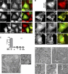
EspF activates SNX9 to form membrane tubules in vivo. (A) Fluorescence microscopy of endogenous SNX9 from EGFP, EGFP-EspF, or EGFP-EspF-D3 transfected HeLa cells. Inset shows the accumulation of SNX9 at CCPs or new tubule structures. Bar = 15 μm. (B) Fluorescence microscopy of HeLa cells transfected with mCherry-SNX9 or the EGFP constructs and mCherry-SNX9 as indicated. Inset shows CCPs (arrows) or new tubule structures. Bar = 15 μm. (C) Quantification of tubule networks in HeLa cells cotransfected with the indicated plasmids. Graphs represent the percentage of transfected cells with tubule networks from at least three independent experiments and SEM is shown. Wild-type (Wt), mutant EspF-D3 (D3), and SNX9ΔBAR (ΔBAR) are indicated. (D) Immunoelectron micrograph of EGFP-EspF in HeLa cells cotransfected with mCherry-SNX9. Sections were labeled with anti-GFP polyclonal antibody and 10 nm protein A-gold. Bars for large image (250 nm) and magnification (50 nm) are indicated. (E–I) Thin-section electron micrograph of EspF- and SNX9-transfected HeLa cells containing multiple membrane tubules showing irregular shapes (arrows) (E). Untransfected controls (F) and representative examples of unusual membrane tubules in EspF transfected cells are shown (G–I). Bars: 500 nm (E and F), 200 nm (G–I).
Figure 6.
Figure 6.
EspF induces N-WASP activation at membrane tubules. (A and B) Fluorescence microscopy images depicting mCherry-SNX9 and FITC-phalloidin (actin) in EspF (A) or EspF-D3 (B) transfected HeLa cells. Bar = 15 μm. (C and D) Fluorescence microscopy images of HeLa cells cotransfected with EGFP-EspF (C) or EGFP-EspF-D3 (D) with mCherry-SNX9 and N-WASP-V5. N-WASP was detected by anti-V5 immunocytochemistry. Bar = 15 μm. (E) Time series of EspF puncta (EGFP-EspF) exhibiting a “rocketing” phenotype (arrow) from live cell TIR-FM. Cartoon below. (F) Time series of EspF puncta (EGFP-EspF) exhibiting a mobile phenotype from live cell TIR-FM. Images are correlated with simultaneous Clc-DsRed dynamics showing the movement of a single CCP (arrow). (G) Schematic of EspF functioning as a node of signaling integration at eukaryotic membranes.
Figure 7.
Figure 7.
SNX9 is the major binding partner for EspF in polarized epithelial cells. (A) TER measurements of polarized T84 colonic epithelial cells infected with EPEC and the indicated EspF mutants. The average change in TER in three independent experiments is shown. (B and C) Fluorescence microscopy of MDCK cells uninfected (B) or infected with EPEC for 4 h (C). Tight junction morphology was detected by anti-occludin immunocytochemistry. (D) Schematic of TAP tagged EspF is shown. Western blot of cellular lysates collected from stable MDCK cells expressing TAP-flag-EspF or mutant TAP-flag-EspF-D3. Anti-flag was used to probe EspF expression (top blot) and actin was used as a protein loading control (bottom blot). (E and F) Fluorescence microscopy of MDCK cells expressing TAP-EspF and TAP-EspF-D3. Tight junction morphology was detected by anti-occludin immunocytochemistry. (G) MDCK parental or Tap-EspF cellular lysates were incubated with anti-flag agarose and the resulting immuno-complexes were subjected to SDS-PAGE and stained with Coomassie. Proteins identified by mass spectrometry are indicated. “NS” designates nonspecific interacting proteins and “IgG LC” is the immunoglobulin light chain. (H) Anti-SNX9 immunofluorescence microscopy of CaCo2 cells infected with the indicated EPEC strains for 3 h. Boxed area is a 4× magnification of the area indicated. Cellular borders are outlined. Bar = 15 μm. (I) Immunoblot of EspF from wild-type EPEC (Wt) or EPEC espF strains carrying plasmids encoding wild-type EspF (pespf) or mutant EspF-D3 (pespf-D3) tagged with the myc epitope. Type III secreted EspF harvested from media supernatants (top) and from whole bacterial lysates (bottom) are shown. Concentrations of IPTG are indicated below.

Similar articles

Cited by

References

    1. Alto, N.M., F. Shao, C.S. Lazar, R.L. Brost, G. Chua, S. Mattoo, S.A. McMahon, P. Ghosh, T.R. Hughes, C. Boone, and J.E. Dixon. 2006. Identification of a bacterial type III effector family with G protein mimicry functions. Cell. 124:133–145. - PubMed
    1. Badour, K., M.K. McGavin, J. Zhang, S. Freeman, C. Vieira, D. Filipp, M. Julius, G.B. Mills, and K.A. Siminovitch. 2007. Interaction of the Wiskott-Aldrich syndrome protein with sorting nexin 9 is required for CD28 endocytosis and cosignaling in T cells. Proc. Natl. Acad. Sci. USA. 104:1593–1598. - PMC - PubMed
    1. Benesch, S., S. Polo, F.P. Lai, K.I. Anderson, T.E. Stradal, J. Wehland, and K. Rottner. 2005. N-WASP deficiency impairs EGF internalization and actin assembly at clathrin-coated pits. J. Cell Sci. 118:3103–3115. - PubMed
    1. Campellone, K.G., D. Robbins, and J.M. Leong. 2004. EspFU is a translocated EHEC effector that interacts with Tir and N-WASP and promotes Nck-independent actin assembly. Dev. Cell. 7:217–228. - PubMed
    1. Co, C., D.T. Wong, S. Gierke, V. Chang, and J. Taunton. 2007. Mechanism of actin network attachment to moving membranes: barbed end capture by N-WASP WH2 domains. Cell. 128:901–913. - PMC - PubMed

Publication types

MeSH terms