. 2007 Dec;27(23):8271-83.
doi: 10.1128/MCB.01781-06. Epub 2007 Sep 24.

Src-dependent phosphorylation of ASAP1 regulates podosomes

Affiliations

Src-dependent phosphorylation of ASAP1 regulates podosomes

Sanita Bharti et al. Mol Cell Biol. 2007 Dec.

Abstract

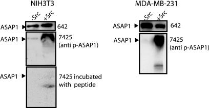
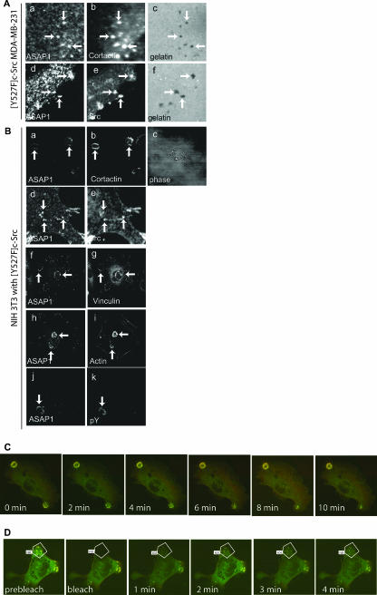
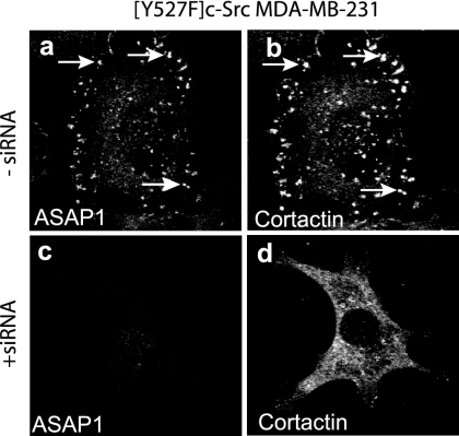
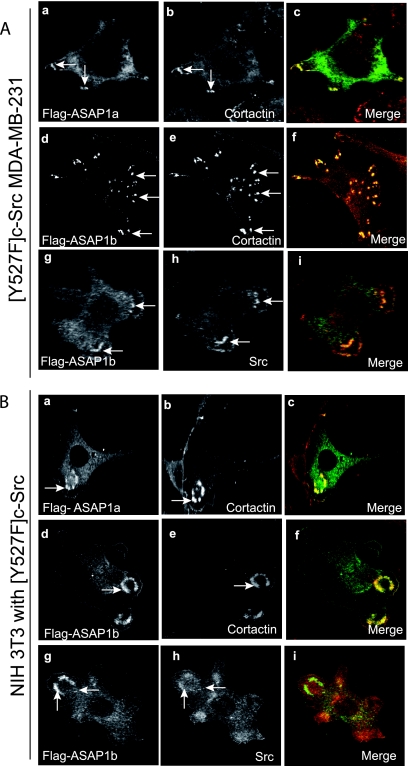
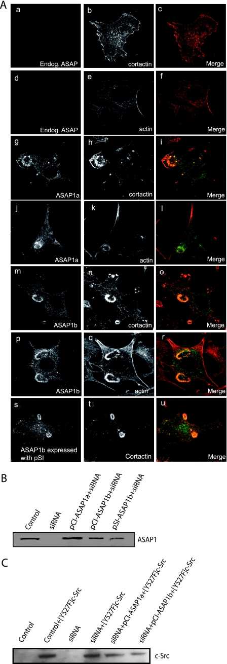
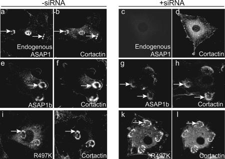
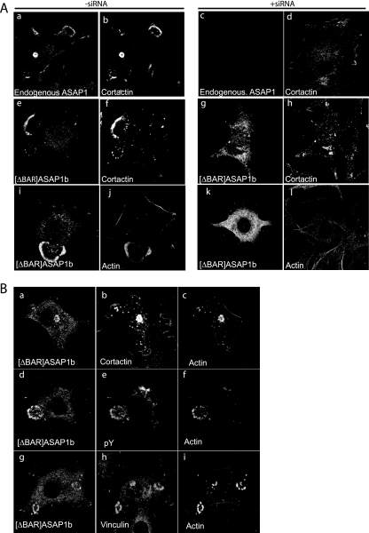
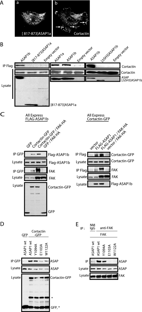
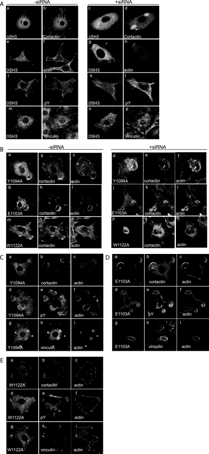
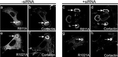
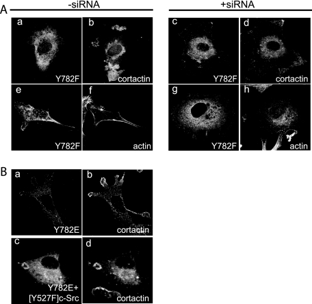
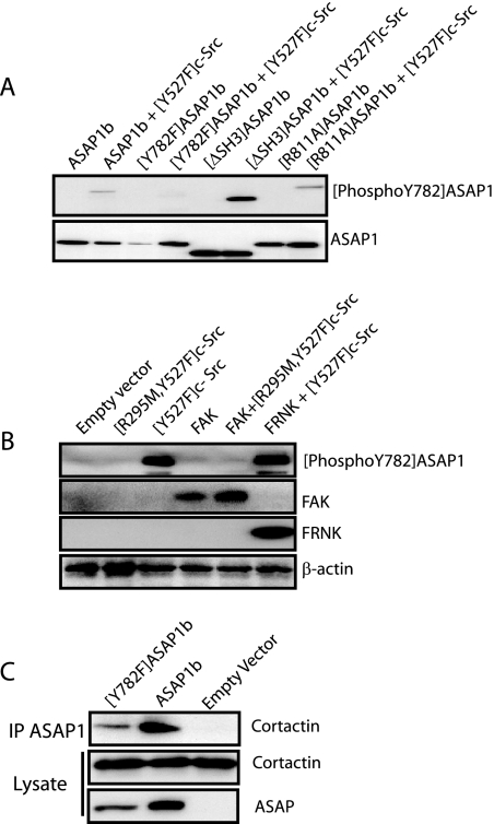
Invadopodia are Src-induced cellular structures that are thought to mediate tumor invasion. ASAP1, an Arf GTPase-activating protein (GAP) containing Src homology 3 (SH3) and Bin, amphiphysin, and RVS161/167 (BAR) domains, is a substrate of Src that controls invadopodia. We have examined the structural requirements for ASAP1-dependent formation of invadopodia and related structures in NIH 3T3 fibroblasts called podosomes. We found that both predominant splice variants of ASAP1 (ASAP1a and ASAP1b) associated with invadopodia and podosomes. Podosomes were highly dynamic, with rapid turnover of both ASAP1 and actin. Reduction of ASAP1 levels by small interfering RNA blocked formation of invadopodia and podosomes. Podosomes were formed in NIH 3T3 fibroblasts in which endogenous ASAP1 was replaced with either recombinant ASAP1a or ASAP1b. ASAP1 mutants that lacked the Src binding site or GAP activity functioned as well as wild-type ASAP1 in the formation of podosomes. Recombinant ASAP1 lacking the BAR domain, the SH3 domain, or the Src phosphorylation site did not support podosome formation. Based on these results, we conclude that ASAP1 is a critical target of tyrosine kinase signaling involved in the regulation of podosomes and invadopodia and speculate that ASAP1 may function as a coincidence detector of simultaneous protein association through the ASAP1 SH3 domain and phosphorylation by Src.


Figures

FIG. 1.
FIG. 1.
Characterization of an antibody raised against a phosphopeptide from ASAP1. An antibody (642) raised against ASAP1 and an affinity-purified antibody (pAB7425) from a serum raised against a phosphopeptide from ASAP1, containing phospho-Y782 (P-Y782), were used to immunoblot lysates from NIH 3T3 and MDA-MB-231 cells expressing [Y527F]c-Src, as indicated. In the lower left panel, pAB7425 was mixed with the peptide against which it was raised prior to immunoblotting.
FIG. 2.
FIG. 2.
Localization of ASAP1. (A) MDA-MB-231 cells. MDA-MB-231 cells stably expressing [Y527F]c-Src were grown on Alexa 488-conjugated gelatin and stained for ASAP1 (a and d) and either cortactin (b) or Src (e). The fluorescently labeled gelatin matrix is visualized (c and f). Black areas (arrows) are regions where gelatin matrix degradation has occurred. (B) NIH 3T3 fibroblasts. NIH 3T3 fibroblasts transiently transfected with plasmids directing the expression of [Y527F]c-Src were grown on a gelatin matrix, fixed, and stained for ASAP1 (a, d, f, h, and j) and either cortactin (b), Src (e), vinculin (g), actin (i), or phosphotyrosine (pY) (k). A phase-contrast image is shown (c). (C) Live-cell imaging of podosomes. NIH 3T3 fibroblasts stably expressing RFP-actin were transfected with plasmids directing the expression of eGFP-ASAP1b and [Y527F]c-Src. The cells were imaged for 10 min. (D) ASAP1 and actin turnover in podosomes. The indicated region of NIH 3T3 fibroblasts were photobleached (area outlined in white), and the recovery of red (RFP-actin) and green (GFP-ASAP1b) fluorescence was determined. roi, region of interest.
FIG. 3.
FIG. 3.
Effect of reduction of ASAP1 levels on [Y527F]c-Src-MDA-MB-231 cells. Cells grown on fibronectin-coated coverslips were treated with siRNA targeting ASAP1 and stained for ASAP1 (a and c) and cortactin (b and d).
FIG. 4.
FIG. 4.
Comparison of ASAP1a and ASAP1b. (A) Localization of ASAP1a and ASAP1b in [Y527F]c-Src-MDA-MB-231 cells. Cells were transfected with plasmids directing expression of Flag-ASAP1a (a to c) and Flag-ASAP1b (d to i). ASAP1 was detected with antibody to the Flag epitope (a, d, and g) and either cortactin (b and e) or Src (h). (B) Localization of ASAP1a and ASAP1b in Src-transformed NIH 3T3 fibroblasts. NIH 3T3 fibroblasts expressing [Y527F]c-Src and either Flag-ASAP1a (a to c) or Flag-ASAP1b (d to i) were stained for ASAP1 using an antibody to the Flag epitope (a, d, and g) and either cortactin (b and e) or Src (h). Panels c, f, and i are the merged images.
FIG. 5.
FIG. 5.
Effect of ASAP1a and ASAP1b on the localization of cortactin in NIH 3T3 fibroblasts transformed with [Y527F]c-Src. NIH 3T3 fibroblasts were transfected with a plasmid directing the expression of [Y527F]c-Src and siRNA to the 3′UTR of ASAP1 to reduce the levels of endogenous ASAP1. As indicated, the cells were also transfected with plasmids with the cytomegalovirus promoter directing the expression of Flag-ASAP1a (pCI-ASAP1a), Flag-ASAP1b (pCI-ASAP1b), or a plasmid with a simian virus 40 promoter directing the expression of Flag-ASAP1b (pSI-ASAP1b); these cDNAs are resistant to the siRNA. (A) Representative micrographs. Podosomes were visualized using an antibody to cortactin or rhodamine-phalloidin. Quantitation is provided in Table 1. (B) Relative expression levels of ASAP1. ASAP1 was detected in the indicated cell lysates by immunoblotting with antibody 642 for ASAP1. (C) Relative expression levels of [Y527F]c-Src. Src in the indicated cell lysates was detected by immunoblotting.
FIG. 6.
FIG. 6.
Effect of ASAP1 Arf GAP activity on podosome formation. The left micrographs (−siRNA) are NIH 3T3 fibroblasts either transfected with a plasmid directing expression of [Y527F]c-Src (a and b) or cotransfected with the plasmid for [Y527F]c-Src and plasmids directing expression of Flag-ASAP1b (e and f) or Flag-[R497K]ASAP1b (i and j). On the right (+siRNA), NIH 3T3 fibroblasts were transfected with plasmids for [Y527F]c-Src and siRNA targeted to the 3′UTR of mouse ASAP1 mRNA and, for some panels, ASAP1b (g and h) or [R497K]ASAP1b (k and l) plasmids resistant to this siRNA. Podosomes were detected with an antibody to cortactin.
FIG. 7.
FIG. 7.
(A) Role of the BAR domain of ASAP1 in podosome formation. The experiment was performed as described for Fig. 6 but using [ΔBAR]ASAP1b to replace endogenous ASAP1. Podosomes were detected with antibodies to cortactin (b, f, d, and h). Cells were also stained for polymerized actin (j and l). (B) Analysis of podosome-like structures formed in the presence of [ΔBAR]ASAP1 and endogenous ASAP1. NIH 3T3 fibroblasts forming podosomes were triple labeled for the indicated proteins or the protein phosphotyrosine (pY).
FIG. 8.
FIG. 8.
ASAP1-cortactin association. (A) Cellular distribution and effects of [817-873]ASAP1a. Flag-[817-873]ASAP1 was expressed in NIH 3T3 fibroblasts with [Y527F]c-Src. Podosomes were detected with an antibody to cortactin. Arrows indicate podosomes. (B) Association of ASAP1 with cortactin. NIH 3T3 fibroblasts were transfected with plasmids directing the expression of the indicated proteins. Association of recombinant ASAP1 with cortactin was determined by immunoprecipitation (IP) through the Flag tag and immunoblotting for both the Flag tag and cortactin. (C) Effect of FAK on ASAP1-cortactin association. Flag-ASAP1b, GFP-cortactin, and HA-FAK were expressed in HEK 293T cells as indicated. In the experimental results shown on the left, proteins were immunoprecipitated with an antibody for GFP. The levels of Flag-ASAP1, GFP-cortactin, and HA-FAK were determined in the lysates and precipitates. In the experimental results shown on the right, proteins were immunoprecipitated with an antibody to the Flag tag. (D) Effect of point mutations within the SH3 domain of ASAP1 on cortactin binding. NIH 3T3 fibroblasts were transfected with expression vectors for Flag-ASAP1 wild type (wt) or the indicated SH3 mutants (W1122A, Y1094A, and E1103A) with GFP-tagged cortactin. Immunoprecipitation was done with anti-GFP polyclonal antibody, and the precipitated materials were analyzed by immunoblotting with monoclonal anti-Flag antibody (M5) or monoclonal anti-GFP antibody. *, GFP-cortactin degradation products. (E) Effect of point mutations within the SH3 domain of ASAP1 on FAK binding. NIH 3T3 fibroblasts were transfected with expression vectors for FLAG-tagged wild-type ASAP1 or the indicated ASAP1 mutants with FAK. Cells were lysed and proteins immunoprecipitated as described for panel D, except that anti-FAK 2A7 monoclonal antibody and normal mouse IgG (NMIgG) (as the negative control) were used instead of anti-GFP for immunoprecipitation.
FIG. 9.
FIG. 9.
(A) Role of SH3 domain of ASAP1 for podosome formation. The experiment was performed as described in the legend to Fig. 6. Representative micrographs are shown, with the quantitation presented in Table 1. Flag-[ΔSH3]ASAP1 transfected cells were detected with polyclonal anti-Flag antibody (a, e, c, and g). Podosomes were detected with cortactin (b and d), actin (f and h), phosphotyrosine (pY) (j and l), and vinculin (n and p). (B) Effect of point mutations within the SH3 domain on podosome formation. NIH 3T3 cells were transfected with the W1122A, Y1094A, and E1103A point mutants and [Y527F]c-Src in the presence (−siRNA) or absence (+siRNA) of endogenous ASAP1. Cells were stained with polyclonal anti-Flag antibody (a, g, m, d, j, and p) and anticortactin antibody (b, h, n, e, k, and q) and for polymerized actin (c, i, o, f, l, and r). (C, D, and E) Analysis of podosome-like structures formed in cells expressing ASAP1 with mutations in the SH3 domain. Cells expressing [Y1094A]ASAP1 (C), [E1103A]ASAP1 (D), or [W1122A]ASAP1 (E) were triple stained for the indicated proteins.
FIG. 10.
FIG. 10.
Role of SH3-binding proline-rich motifs in ASAP1 for podosome formation. The experiment was performed as described in the legend to Fig. 6 using plasmids directing expression of Flag-[R811A]ASAP1b or Flag-[R1021A]ASAP1b. Quantitation is provided in Table 1. Transfected cells were visualized using polyclonal anti-Flag antibody (a, e, c, and g). Podosomes were visualized using cortactin (b, f, d, and h).
FIG. 11.
FIG. 11.
Role of Tyr-782 of ASAP1 for podosome formation. (A) [Y782F]ASAP1. This experiment was performed as described in the legend to Fig. 6 using a plasmid directing expression of [Y782F]ASAP1. Quantitation is provided in Table 1. (B) Localization of [Y782E]ASAP1. Flag-[Y782E]ASAP1 was expressed in NIH 3T3 fibroblasts that also expressed [Y527F]c-Src where indicated (c). The cells were immunostained with antibodies against the Flag epitope (a and c) and cortactin (b and d).
FIG. 12.
FIG. 12.
Src-dependent phosphorylation of ASAP1. (A) Phosphorylation of ASAP1 mutants. Phosphorylation of Flag-ASAP1, Flag-[Y782F]ASAP1, Flag-[ΔSH3]ASAP1, and Flag-[R811A]ASAP1 expressed in NIH 3T3 fibroblasts with [Y527F]c-Src, as indicated, was determined by immunoblotting with anti-[P-Y782]ASAP1 antibody, pAB7425 (see Materials and Methods for a description of this antibody). Total cell lysates were also probed with monoclonal Flag antibody to control for transfection efficiency (lower panel). (B) Role of Src and FAK in phosphorylation of ASAP1. [P-Y782]ASAP1 was determined in lysates of NIH 3T3 fibroblasts expressing [R295M, Y527F]c-Src, [Y527F]c-Src, FAK, FAK with [R295M, Y527F]c-Src, or [Y527F]c-Src with FRNK. Total cell lysates were prepared and probed with anti-[P-Y782]ASAP1 (antibody pAB7425), anti-FAK, anti-myc for detection of myc-FRNK, and an antibody against actin to control for loading. (C) Role of phosphorylation in cortactin binding. The indicated recombinant Flag-ASAP1b protein was expressed in NIH 3T3 fibroblasts. Proteins were immunoprecipitated through the Flag epitope. Immunoblottings of the immunoprecipitated (IP) material and total cell lysates were performed as indicated.

References

    1. Arold, S., P. Franken, M. P. Strub, F. Hoh, S. Benichou, R. Benarous, and C. Dumas. 1997. The crystal structure of HIV-1 Nef protein bound to the Fyn kinase SH3 domain suggests a role for this complex in altered T cell receptor signaling. Structure 5:1361-1372. - PubMed
    1. Bowden, E. T., M. Barth, D. Thomas, R. I. Glazer, and S. C. Mueller. 1999. An invasion-related complex of cortactin, paxillin and PKCμ associates with invadopodia at sites of extracellular matrix degradation. Oncogene 18:4440-4449. - PubMed
    1. Brown, M. T., J. Andrade, H. Radhakrishna, J. G. Donaldson, J. A. Cooper, and P. A. Randazzo. 1998. ASAP1, a phospholipid-dependent Arf GTPase-activating protein that associates with and is phosphorylated by Src. Mol. Cell. Biol. 18:7038-7051. - PMC - PubMed
    1. Brunton, V. G., I. R. J. MacPherson, and M. C. Frame. 2004. Cell adhesion receptors, tyrosine kinases and actin modulators: a complex three-way circuitry. Biochim. Biophys. Acta 1692:121-144. - PubMed
    1. Buccione, R., J. D. Orth, and M. A. McNiven. 2004. Foot and mouth: podosomes, invadopodia and circular dorsal ruffles. Nat. Rev. Mol. Cell Biol. 5:647-657. - PubMed

Publication types

MeSH terms