. 2007 Dec;27(23):8340-51.
doi: 10.1128/MCB.00972-07. Epub 2007 Sep 24.

ZBP2 facilitates binding of ZBP1 to beta-actin mRNA during transcription

Affiliations

ZBP2 facilitates binding of ZBP1 to beta-actin mRNA during transcription

Feng Pan et al. Mol Cell Biol. 2007 Dec.

Erratum in

  • Mol Cell Biol. 2009 May;29(9):2481

Abstract

Cytoplasmic mRNA localization regulates gene expression by spatially restricting protein translation. Recent evidence has shown that nuclear proteins (such as hnRNPs) are required to form mRNPs capable of cytoplasmic localization. ZBP1 and ZBP2, two hnRNP K homology domain-containing proteins, were previously identified by their binding to the zipcode, the sequence element necessary and sufficient for beta-actin mRNA localization. ZBP1 colocalizes with nascent beta-actin mRNA in the nucleus but is predominantly a cytoplasmic protein. ZBP2, in contrast, is predominantly nuclear. We hypothesized that the two proteins cooperate to localize beta-actin mRNA and sought to address where and how this might occur. We demonstrate that ZBP2, a homologue of the splicing factor KSRP, binds initially to nascent beta-actin transcripts and facilitates the subsequent binding of the shuttling ZBP1. ZBP1 then associates with the RNA throughout the nuclear export and cytoplasmic localization process.


Figures

FIG. 1.
FIG. 1.
ZBP2 binds to the chicken β-actin zipcode with all four KH domains. (A) Purification of ZBP2 from SF9 insect cells. His-tagged ZBP2 (lane E) was purified from insect cell lysates (lane L) after three washes (lanes W1 to W3). Ld is benchmark protein ladder (Invitrogen). (B) Three ZBP2 truncations, i.e., the first and second KH domains (KH1-2), the third and fourth KH domains (KH3-4), and all four KH domains (KH1-4), were expressed and purified using the same system as for panel A. Ld indicates a 10-kDa protein ladder (Invitrogen). (C) Gel shift with 32P-labeled chicken β-actin zipcode probe using various truncations of ZBP2 KH domains. The full-length ZBP2-zipcode complex is indicated by an arrow. All four KH domains of ZBP2 were required for efficient binding. (D) Nitrocellulose filter binding assay of the ZBP2 fragments in panel C, showing affinity curves for the β-actin mRNA zipcode. Recombinant full-length ZBP2 and three fragments, i.e., KH1-2, KH3-4, and KH1-4, at various concentrations were incubated with 32P-labeled zipcode RNA probe. Bound probe was detected by Cerenkov counting after binding of the protein-RNA complex to the filter and intensive washing.
FIG. 2.
FIG. 2.
ZBP2 binds to pyrimidine-rich RNA ligands. (A) SELEX was used to select RNA motifs specifically recognized by ZBP2. After 10 rounds of selection and amplification, 20 clones were sequenced, and two groups with conserved patterns are aligned and underlined: the CCCC motif and the GUCC motif. (B) RNA competitor sequences used for panels C and D. The zipcode was in vitro transcribed as described in Materials and Methods. SELEX competitors were in vitro transcribed from PCR products amplified from SELEX clones S1 (ZBP1 SELEX), S2, and S3 by T7 polymerase. (C) RNA competition shows that the two SELEX oligonucleotides efficiently competed with the zipcode for ZBP2 binding. No competitor or 100× concentrations of different competitors were preincubated with ZBP2 for 15 min. 32P-labeled chicken β-actin zipcode probe was added to the mix and incubated for a further 30 min. The RNA-protein complexes were resolved in a 6% native gel. The arrow indicates the position of the ZBP2-zipcode complexes. (D) Binding of the zipcode to ZBP1 was not competed with the CCCC or GUCC motif. RNA competition assays were performed as for panel C. The position of the ZBP1-zipcode complexes is indicated.
FIG. 3.
FIG. 3.
Recombinant ZBP2 and ZBP1 do not simultaneously bind to the zipcode. (A) RNA footprinting assays using V1 nuclease digestion to determine the binding sites for ZBP1 and ZBP2 on the 54-nt zipcode of chicken β-actin mRNA. ZBP2 increased RNase V1 digestion efficiency in the ZBP1 binding region, resulting in stronger bands (arrowheads and torpedo) as well as a moderate protection of additional bands (arrow). (B) The 32P-labeled chicken 56-nt zipcode was incubated with recombinant ZBP1 KH1-4, ZBP2, or both proteins and then resolved on a 6% native gel. ZBP1 KH1-4 bound to the probe most likely as a monomer (arrow), with minor signal above, presumably ZBP1 dimers. The results indicated that in solution, when both ZBP1 and ZBP2 were present, the zipcode preferentially bound to recombinant ZBP2 (arrowhead).
FIG. 4.
FIG. 4.
ZBP2 binds to and colocalizes with nascent β-actin mRNA. (A) MTC cells, which were serum responsive, were either serum starved overnight or stimulated with serum for 15 min. The cells were subjected to ChIP using anti-acetylated histone antibody, normal rat IgG, or polyclonal antibodies against the C terminus of mZBP2 (a generous gift from Doug Black). Two primer pairs, one amplifying the 3′ end of the β-actin-coding region and the other amplifying the 5′ end, which was approximately 3 kb upstream of stop codon, were used in PCR experiments after ChIP. mZBP2 was associated only with the 3′ end of the β-actin-coding region after serum stimulation (A, upper panel, arrow) and not with the 5′ end (A, lower panel, arrow). (B to E) ZBP2 and ZBP1 colocalize at the β-actin mRNA transcription site. Yellow fluorescent protein-ZBP1 (green) (B) and cyan fluorescent protein-mZBP2 (blue) (D) were cotransfected overnight into NG108 cells. Cells were plated on Cell-Tek (BD Bioscience)-coated coverslips and then serum starved for 12 h for differentiation. Nuclear foci were observed with colocalization (arrow) of ZBP1 (B), ZBP2 (D), and β-actin mRNA (red) (C). The merged image is shown in panel E. Bar, 10 μm.
FIG. 5.
FIG. 5.
Efficient knockdown of ZBP2 by RNAi. (A) RNAi effect caused by ZBP2 siRNAs in 293 cells. siRNA oligonucleotides against the KH domain or the N terminus of ZBP2/KSRP were transfected overnight into HEK293 cells with Lipofectamine 2000. Cells were aliquoted to six-well plates, and samples were collected for whole-cell lysates and total RNA preparation at 24, 48, 72, and 96 h after plating (D1 to -4). A monoclonal antibody was used in the Western blot to detect KSRP/ZBP2 expression, whereas vinculin was used as loading control. (B) ZBP2 mRNA was degraded by siRNA against the KH domain of KSRP/ZBP2. A scrambled siRNA (S) N-terminal ZBP2 siRNA (N), or KH domain ZBP2 siRNA (KH) was transfected into 293 cells as described above, and total RNA was isolated, reverse transcribed, and subjected to real-time PCR. ZBP1 (black) and ZBP2 (white) relative mRNA levels were normalized and averaged from the values from three independent experiments. Error bars indicate standard deviations. (C) Colocalization of ZBP1 and β-actin mRNA at the transcription sites requires ZBP2. NG108-15 cells were transfected with ZBP2 KH domain siRNA and differentiated as described in Materials and Methods. After in situ hybridization against β-actin mRNA, the percentage of cells with ZBP1 colocalized with transcription sites was counted in cells with ZBP2 (ZBP2+) or without ZBP2 (ZBP2−) after RNAi (P < 0.001).
FIG. 6.
FIG. 6.
ZBP1 recruitment to β-actin transcription sites is ZBP2 dependent. (A) Time course of ZBP1 and ZBP2 recruitment. CEFs synchronized by serum starvation were serum stimulated and then fixed at 5-min intervals. At least 200 cells with ZBP1 and ZBP2 signals were counted at each time point. The diagram shows the average percentage of cells with ZBP1 or ZBP2 colocalized with β-actin nascent transcripts from two independent experiments. Error bars indicate standard deviations. (B) ZBP2 is required for efficient binding of ZBP1 to nascent β-actin mRNA. MTC cells were transfected with either scrambled RNAi oligonucleotide (lanes 1 to 4) or ZBP2 RNAi oligonucleotide (lanes 6 to 9) for 2 days and subjected to ChIP. Two primer pairs were used to amplify the 3′ coding regions of the β-actin (top) and GAPDH (bottom) genes from the precipitates. Lanes 1 and 6, inputs of total nuclear lysates; lanes 3 and 8, anti-acetylated histone antibody was used as positive control; lanes 2 and 7, normal rat IgG was used as negative control. Lanes 4 and 9 used polyclonal antibodies against chicken ZBP1. Binding of the β-actin gene with acetylated histone was unaffected in the ZBP2 knockdown cells (compare lane 3 to lane 8). However, knockdown of ZBP2 expression reduced the binding of ZBP1 to nascent β-actin mRNA by 65% (compare lane 4 to lane 9). (C) Chromatin lysates from cultured MTC cells were treated with RNase A and subjected to precipitation with antibodies against histone (lanes 3 and 4) and ZBP1 (lanes 5 and 6). The precipitation efficiency of ZBP1 with the β-actin gene was drastically affected in the RNase A-treated cell lysates, indicating that their association is transcription dependent. Lane 2 indicates the lysate input. (D) Effective binding of ZBP1 to the zipcode requires ZBP2. CEF extracts were preincubated with 100× unlabeled zipcode and different amounts of CCCC' containing SELEX2 oligonucleotides for 15 min. 32P-labeled chicken β-actin zipcode probe was added to the mixes and incubated for additional 30 min. The RNA-protein complexes were separated in a 6% native gel. The arrows point the positions of the three zipcode complexes (S and M, ZBP2 complexes; F, ZBP1 complex).
FIG. 7.
FIG. 7.
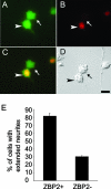
ZBP2 is required for neurite outgrowth. (A to D) Knockdown of ZBP2 inhibits neurite outgrowth. Differentiating NG108-15 cells were cotransfected with a GFP-expressing plasmid and with either scrambled siRNA or ZBP2 KH domain siRNA for 48 h. The two populations were mixed and plated on Cell-Tek-coated coverslips. Cells were seeded overnight and then subjected to 12 h of differentiation and immunofluorescence detection against ZBP2. GFP (A) (green), ZBP2 (B) (red), merged (C), and differential interference contrast (D) images are shown. ZBP2 knockdown cells (arrows) did not form elongated neurites, in contrast to ZBP2-positive cells (arrowheads). Bar, 10 μm. (E) Statistical analysis of effects of ZBP2 knockdown on neurite growth. More than 80% of the ZBP2-positive (ZBP+) cells have neurites longer than 1.5 times the cell diameter, while only 33% of ZBP2 knockdown (ZBP−) cells have elongated neurites. At least 200 cells were counted in each of three independent experiments. Error bars indicate standard deviations.

Similar articles

Cited by

References

    1. Adams, M. D., R. S. Tarng, and D. C. Rio. 1997. The alternative splicing factor PSI regulates P-element third intron splicing in vivo. Genes. Dev. 11:129-138. - PubMed
    1. Amarasinghe, A. K., R. MacDiarmid, M. D. Adams, and D. C. Rio. 2001. An in vitro-selected RNA-binding site for the KH domain protein PSI acts as a splicing inhibitor element. RNA 7:1239-1253. - PMC - PubMed
    1. Bassell, G. J., and R. H. Singer. 2001. Neuronal RNA localization and the cytoskeleton. Probl. Cell Differ. 34:41-56. - PubMed
    1. Bassell, G. J., H. Zhang, A. L. Byrd, A. M. Femino, R. H. Singer, K. L. Taneja, L. M. Lifshitz, I. M. Herman, and K. S. Kosik. 1998. Sorting of beta-actin mRNA and protein to neurites and growth cones in culture. J. Neurosci. 18:251-265. - PMC - PubMed
    1. Carey, J., V. Cameron, P. L. de Haseth, and O. C. Uhlenbeck. 1983. Sequence-specific interaction of R17 coat protein with its ribonucleic acid binding site. Biochemistry 22:2601-2610. - PubMed

Publication types

MeSH terms

LinkOut - more resources