. 2007 Dec;27(23):8243-58.
doi: 10.1128/MCB.00899-07. Epub 2007 Sep 24.

Major and essential role for the DNA methylation mark in mouse embryogenesis and stable association of DNMT1 with newly replicated regions

Affiliations

Major and essential role for the DNA methylation mark in mouse embryogenesis and stable association of DNMT1 with newly replicated regions

Shin-ichiro Takebayashi et al. Mol Cell Biol. 2007 Dec.

Abstract

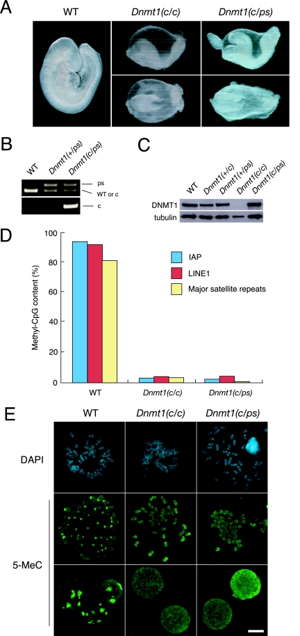
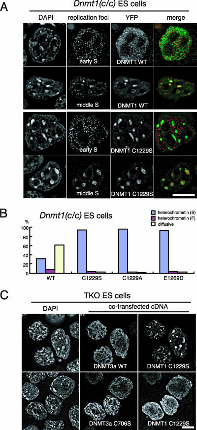
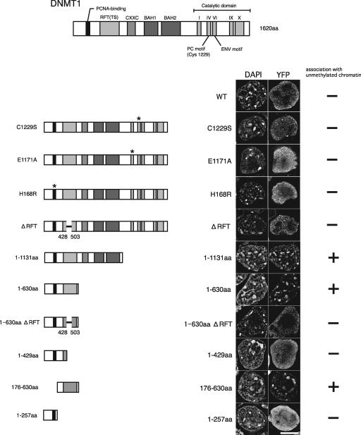
DNA methyltransferase 1 (DNMT1) plays an important role in the inheritance of genomic DNA methylation, which is coupled to the DNA replication process. Early embryonic lethality in DNMT1-null mutant (Dnmt1(c)) mice indicates that DNA methylation is essential for mammalian development. DNMT1, however, interacts with a number of transcriptional regulators and has a transcriptional repressor activity independent of its catalytic activity. To examine the roles of the catalytic activity of DNMT1 in vivo, we generated a Dnmt1(ps) allele that expresses a point-mutated protein that lacks catalytic activity (DNMT1-C1229S). Dnmt1(ps) mutant mice showed developmental arrest shortly after gastrulation, near-complete loss of DNA methylation, and an altered distribution of repressive chromatin markers in the nuclei; these phenotypes are quite similar to those of the Dnmt1(c) mutant. The mutant DNMT1 protein failed to associate with replication foci in Dnmt1(ps) cells. Reconstitution experiments and replication labeling in Dnmt1-/- Dnmt3a-/- Dnmt3b-/- (i.e., unmethylated) embryonic stem cells revealed that preexisting DNA methylation is a major determinant for the cell cycle-dependent localization of DNMT1. The C-terminal catalytic domain of DNMT1 inhibited its stable association with unmethylated chromatin. Our results reveal essential roles for the DNA methylation mark in mammalian development and in DNMT1 localization.


Figures

FIG. 1.
FIG. 1.
Generation of the Dnmt1ps allele by homologous recombination in ES cells. (A) Schematic illustration of the DNMT1 structure. (B) Generation of the Dnmt1-ps allele. The wild-type Dnmt1 locus was targeted with pTS015 carrying a mutated exon 32 and the IRES-βgeo cassette with flanking loxP sites (Dnmt1-neo-ps allele). After homologous recombination, the cassette was removed by Cre recombinase, resulting in the Dnmt1-ps allele. Cysteine 1229 of PC residues (box shade) in motif IV was changed to serine. The remaining wild-type locus was targeted by pTA010, in which exons 31 to 33 were deleted, to create a null mutation. Filled boxes indicate exons, a red star indicates a mutated exon, and arrowheads indicate loxP sites. Locations of the 5′ and 3′ probes for genomic Southern analysis are shown. (C) Genomic Southern analysis of the targeted ES cells digested with EcoRV for the 5′ probe (left) or XmaI for the 3′ probe (right). The 5′ probe detects a >20-kb band (wild type, ps) or an 8.8-kb band (neo-ps, null), and the 3′ probe detects a 20-kb band (wild-type, ps), a 14-kb band (neo-ps), or a 12.5-kb band (null). WT, wild-type. (D) Sequences of Dnmt1 transcripts in wild-type and Dnmt1−/ps ES cells. The red star indicates the mutated base. (E) Immunoblot analysis of ES cell extracts by use of an anti-DNMT1 antibody. (F) DNA methylation of repetitive sequences (IAP and pericentromeric major satellite repeats), analyzed by Southern blotting. Genomic DNA from each cell line was digested with methylation-sensitive (HpaII or MaeII) or methylation-insensitive (MspI) restriction enzymes and analyzed with the probe indicated at the bottom of each blot. (G) Quantitative analysis of the methyl-CpG content of repetitive sequences (IAP, LINE1, and pericentromeric major satellite repeats) by bisulfite sequencing analysis. The percentages of methyl-CpG in the total CpG sites analyzed in wild-type and mutant ES cells are indicated. (H) Anti-methylcytosine antibody staining in wild-type, Dnmt1c/c, Dnmt1−/ps, and Dnmt1−/− Dnmt3a−/− Dnmt3b−/− (TKO) ES cells. Mitotic chromosomes were stained with DAPI (blue). Distribution of methyl-cytosine (green) was detected in mitotic chromosomes and interphase nuclei. Scale bar, 10 μm.
FIG. 2.
FIG. 2.
Early embryonic lethality of Dnmt1c/ps embryos. (A) Gross morphology of wild-type (WT), Dnmt1c/c, and Dnmt1c/ps embryos dissected at E9.5. Growth of Dnmt1c/c and Dnmt1c/ps embryos was retarded at similar embryonic stages. Embryos of both mutant genotypes formed no or few (up to seven) somites and had distorted neural tubes. (B) PCR genotype analysis of genomic DNA isolated from wild-type, Dnmt1ps/+, and Dnmt1c/ps embryos. The upper panel shows results for a primer set for the Dnmt1-ps allele, and the lower panel shows results for a primer set for the Dnmt1-c allele (see Materials and Methods). (C) Immunoblot analysis of embryonic cell extracts (E9.5) probed with an anti-DNMT1 antibody. For the loading control, the same membrane was stripped and reprobed with an antitubulin antibody. (D) Bisulfite sequencing analysis of repetitive sequences (IAP, LINE1, and pericentromeric major satellite repeats) as described for Fig. 1G. Genomic DNA was isolated from E9.5 embryos. (E) Anti-methylcytosine antibody staining in wild-type, Dnmt1c/c, and Dnmt1c/ps cells from E9.5 embryos. Mitotic chromosomes were visualized by DAPI staining (blue). Distribution of methyl-cytosine (green) was detected in mitotic chromosomes and interphase nuclei. Scale bar, 10 μm.
FIG. 3.
FIG. 3.
Growth defect and damage response in Dnmt1c/ps embryonic cells. (A) RT-PCR expression analysis of cell cycle-related (p21, p27, and p53) and DNA methylation-sensitive (H19, Igf2, and IAP) genes in wild-type (WT), Dnmt1c/c, and Dnmt1c/ps embryos at E9.5. GAPDH (glyceraldehyde-3-phosphate dehydrogenase) was used as the loading control. (B) Immunoblot analysis of p53 phosphorylated on Ser15 (p53-P). The results for the loading control, β-tubulin, were the same as those in Fig. 2C. (C) Immunofluorescence staining of PCNA in wild-type, Dnmt1c/c, and Dnmt1c/ps embryonic cells. Nuclei were visualized by DAPI staining. Conventional microscopic images are shown. Scale bar, 10 μm. Average percentages of PCNA-positive cells, with standard deviations, are shown at right (n > 300, triplicate). The difference between the wild-type and Dnmt1c/c or Dnmt1c/ps embryonic cells was statistically significant (P < 0.01, chi-square analysis).
FIG. 4.
FIG. 4.
Dysregulation of chromatin structure in Dnmt1c/ps embryos. (A and B) Immunofluorescence staining of embryonic cells isolated from wild-type (WT) and mutant embryos at E9.5 with specific antibodies to HP1β (A) and H3K9me3 (B). Nuclei were stained with DAPI. The upper panels are low-magnification images (conventional microscopic images), and the lower panels are high-magnification images (deconvolution images). Percentages of cells showing the decreased heterochromatin signal (left) and diffuse staining pattern (right) of HP1β are shown (A). For H3K9me3 staining, interphase (left) and mitotic (right) nuclei are shown (B). (C) Immunoblot analysis of HP1β in the Triton-resistant and Triton-extracted fractions of embryonic cells. PCNA was used as the control for the Triton-extracted fraction. (D, E) Immunostaining analysis of acetylated H4 in embryonic cells. A small population of cells in both the Dnmt1c/c and Dnmt1c/ps embryos had abnormally hyperacetylated histone H4 (D). An abnormal association of HP1β proteins with mitotic chromosomes with hyperacetylated histone H4 was observed (E). Scale bars, 10 μm.
FIG. 5.
FIG. 5.
Perturbed localization of DNMT1 in Dnmt1c/ps embryos and Dnmt1−/ps ES cells. Localization of endogenous DNMT1 proteins in wild-type embryos (A to C), wild-type ES cells (D to F), Dnmt1c/ps embryos (G to I), and Dnmt1−/ps ES cells (J to L) is shown. Colocalization of DNMT1 (green) with PCNA (red) was visualized in the early S (A, D, G, and J), middle S (B, E, H, and K), and late S (C, F, I, and L) phases. Merged images at right show overlays of PCNA and DNMT1 staining (yellow indicates overlap). Nuclei were stained with DAPI (blue). (A, D, G, and J) Magnified views of the regions of the early-S-phase cells in white squares are shown. Scale bars, 10 μm.
FIG. 6.
FIG. 6.
Essential role for DNA methylation mark in DNMT1 localization. (A to H) Localization of YFP-DNMT1 fusion proteins during replication of the pericentromeric heterochromatin. YFP fusions with wild-type (A and E) and mutant (C1229S [B and F], H168R [C and G], and ΔRFT [D and H]) DNMT1 proteins were transiently expressed in wild-type (A to D) and TKO (E to H) ES cells. Replication sites were visualized by the incorporation of digoxigenin-dUTP. Nuclei were visualized by DAPI staining. Merged images of YFP-DNMT1 (green) and replication foci (red) are shown. (I to L) YFP fusions with wild-type (I and J) and mutant (H168R [K] and ΔRFT [L]) DNMT1 proteins were transiently coexpressed with wild-type (I, K, and L) or catalytic-defective (J) DNMT3A proteins in TKO ES cells, and the recovery of the cell cycle-dependent localization of DNMT1 was assessed. DNMT3A proteins were detected by immunofluorescence staining. (M) Localization of DNMT1 at replicated regions in Dnmt3a−/− Dnmt3b−/− ES cells. Results are shown for immunofluorescence staining of endogenous DNMT1 and PCNA proteins in low-passaged Dnmt3a−/− Dnmt3b−/− ES cells. Nuclei were visualized by DAPI staining. A merged image of DNMT1 (green) and PCNA (red) is shown. Scale bars, 10 μm.
FIG. 7.
FIG. 7.
Mislocalization of catalytic-defective DNMT1 to heterochromatin in Dnmt1-deficient ES cells in a methylation mark-dependent manner. (A) Localization of YFP-fused wild-type and C1229S DNMT1 proteins during the replication of euchromatin (early S) and pericentromeric heterochromatin (middle S) in Dnmt1c/c ES cells. Replication sites were visualized by the incorporation of digoxigenin-dUTP. Nuclei were visualized by DAPI staining. Merged images of YFP-DNMT1 (green) and replication foci (red) are shown. (B) Localization analysis of YFP-fused wild-type or catalytic-defective DNMT1 in Dnmt1c/c ES cells. DNMT1 localization was classified into three patterns: focal heterochromatin (S; blue), characterized by strong localization to pericentromeric heterochromatin; faint heterochromatin (F; red), characterized by localization to both pericentromeric heterochromatin and euchromatin; and diffuse (yellow), charac-terized by diffuse localization throughout the euchromatin. The percentage of cells showing each staining pattern is shown (n > 180). The catalytic-defective YFP-DNMT1 mutants were retained in the heterochromatin regions in the Dnmt1c/c ES cells. (C) YFP-DNMT1-C1229S fusion protein was transiently coexpressed with the wild-type (WT) or catalytic-defective (C706S) DNMT3A protein in TKO ES cells. DNMT3A proteins were detected by immunofluorescence staining. Scale bars, 10 μm.
FIG. 8.
FIG. 8.
Prompt and stable association of DNMT1-C1229S with hemi-methylated sites introduced by 5-methylcytosine incorporation in TKO ES cells. (A) Summary of the experimental procedure. YFP-DNMT1-C1229S fusion protein was transiently expressed in TKO ES cells, which were then treated with hypotonic buffer for 5 min to introduce methylated or unmethylated dCTP exogenously, cultured for 1 h, and then fixed for immunofluorescence detection. Biotin-dUTP was introduced simultaneously with dCTP and detected with avidin-Cy3 to visualize replication sites in the nucleus. (B) Cell nuclei that incorporated methylated dCTP during euchromatic replication (top) and pericentromeric replication (middle) or incorporated unmethylated dCTP (bottom) during pericentromeric replication are shown. Magnified views (insets) are shown for the sites of pericentromeric replication marked with white squares. Scale bar, 10 μm. (C) YFP-DNMT1-C1229S fusion protein was transiently coexpressed with untagged wild-type (WT) or catalytic-defective DNMT1 in TKO ES cells before methylated-dCTP incorporation. The percentages of YFP-DNMT1-C1229S enrichment at pericentromeric replication sites (biotin-dUTP positive) are shown (n = 50). The number of cells that were labeled with biotin-dUTP at the pericentromeric heterochromatin regions is considered 100%.
FIG. 9.
FIG. 9.
Inhibitory effect of the C-terminal catalytic domain on pericentromeric heterochromatin localization in TKO ES cells. Maps of YFP-DNMT1 deletion mutant constructs are shown at left. Representative images of TKO ES cells expressing each construct are shown. Association with pericentromeric heterochromatin was assessed for each mutant protein. Nuclei were visualized by DAPI staining. WT, wild type. Scale bar, 10 μm.

References

    1. Araujo, F. D., S. Croteau, A. D. Slack, S. Milutinovic, P. Bigey, G. B. Price, M. Zannis-Hajopoulos, and M. Szyf. 2001. The DNMT1 target recognition domain resides in the N terminus. J. Biol. Chem. 276:6930-6936. - PubMed
    1. Bacolla, A., S. Pradhan, R. J. Roberts, and R. D. Wells. 1999. Recombinant human DNA (cytosine-5) methyltransferase. II. Steady-state kinetics reveal allosteric activation by methylated dna. J. Biol. Chem. 274:33011-33019. - PubMed
    1. Bird, A. 2002. DNA methylation patterns and epigenetic memory. Genes Dev. 16:6-21. - PubMed
    1. Carlone, D. L., J. H. Lee, S. R. Young, E. Dobrota, J. S. Butler, J. Ruiz, and D. G. Skalnik. 2005. Reduced genomic cytosine methylation and defective cellular differentiation in embryonic stem cells lacking CpG binding protein. Mol. Cell. Biol. 25:4881-4891. - PMC - PubMed
    1. Chen, T., S. Hevi, F. Gay, N. Tsujimoto, T. He, B. Zhang, Y. Ueda, and E. Li. 2007. Complete inactivation of DNMT1 leads to mitotic catastrophe in human cancer cells. Nat. Genet. 39:391-396. - PubMed

Publication types

MeSH terms

Substances

LinkOut - more resources