

doi: 10.1073/pnas.0707782104.
Epub 2007 Sep 24.
Suppression of p53-dependent senescence by the JNK signal transduction pathway
Affiliations
- PMID: 17893331
- PMCID: PMC2000443
- DOI: 10.1073/pnas.0707782104
Item in Clipboard
Suppression of p53-dependent senescence by the JNK signal transduction pathway
Proc Natl Acad Sci U S A.
.
Abstract
The JNK signaling pathway is implicated in the regulation of the AP1 transcription factor and cell proliferation. Here, we examine the role of JNK by using conditional and chemical genetic alleles of the ubiquitously expressed murine genes that encode the isoforms JNK1 and JNK2. Our analysis demonstrates that JNK is not essential for proliferation. However, JNK is required for expression of the cJun and JunD components of the AP1 transcription factor, and JNK-deficient cells exhibit early p53-dependent senescence. These data demonstrate that JNK can act as a negative regulator of the p53 tumor suppressor.
Conflict of interest statement
The authors declare no conflict of interest.
Figures
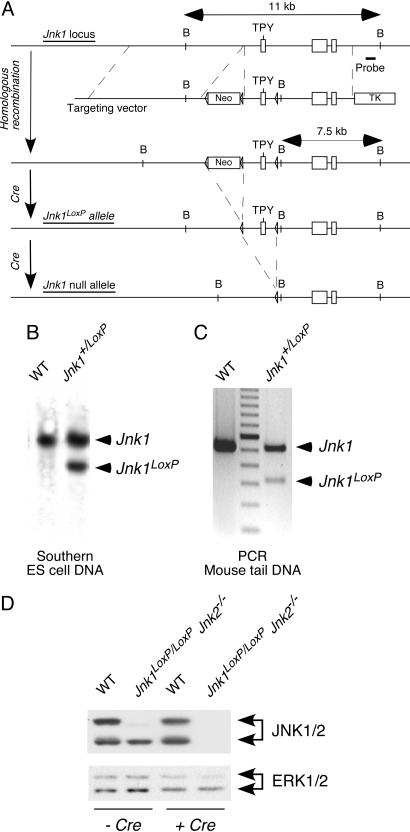
Construction of a conditional Jnk1 allele in mice. (A) The strategy used for targeting the Jnk1 gene to create a conditional Jnk1LoxP allele is illustrated. A probe used for Southern blot analysis and the expected BamH1 restriction fragments are shown. (B) Southern blot analysis of BamH1-restricted genomic DNA isolated from WT and Jnk1+/LoxP ES cells is shown. (C) Genomic DNA was isolated from the tails of WT and Jnk1+/LoxP mice and examined by PCR analysis. (D) Primary MEF were isolated from WT mice and Jnk1LoxP/LoxP Jnk2−/− mice. The MEF were transduced by using a retroviral Cre expression vector. The MEF were cultured (7 days) and extracts were examined by immunoblot analysis using antibodies to JNK1/2 and ERK1/2.
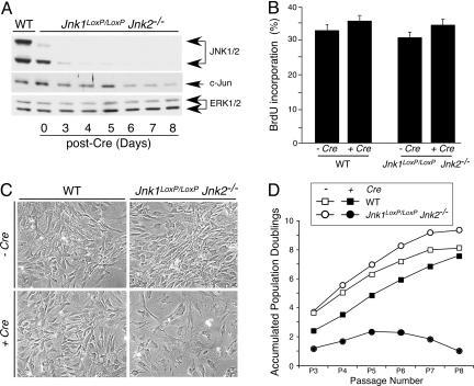
JNK deficiency causes a delayed reduction in cellular proliferation. (A) Time-course analysis of JNK expression after gene ablation. Jnk1LoxP/LoxP Jnk2−/− MEF were transduced by using a retroviral Cre expression vector. Cell extracts were prepared on different days after infection and examined by immunoblot analysis using antibodies to JNK, cJun, and ERK. Extracts prepared from WT MEF were also examined. (B) The effect of retroviral tranduction of Cre in WT and Jnk1LoxP/LoxP Jnk2−/− MEF was examined 10 days after infection by measurement of the incorporation of BrdU by flow cytometry. (C) The morphology of WT and Jnk1LoxP/LoxP Jnk2−/− MEF at passage 7 after retroviral transduction of Cre was examined by phase-contrast microscopy. (Magnification: ×100.) (D) The growth of WT and Jnk1LoxP/LoxP Jnk2−/− MEF was examined by using a 3T3 assay. The data are presented as the accumulated population doublings during eight passages in culture. The effect of retroviral transduction of Cre was investigated.
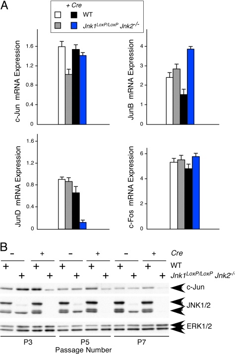
JNK deficiency causes reduced expression of cJun and JunD. (A) WT and Jnk1LoxP/LoxP Jnk2−/− MEF were transduced by using a Cre retroviral expression vector. RNA was isolated from these cells at passage 7 postinfection and examined by quantitative RT-PCR (Taqman) analysis using probes for AP-1 transcription factors (cJun, JunB, JunD, and cFos) and GAPDH. The data are presented as the normalized ratio of [AP-1 mRNA]/[GAPDH mRNA]. (B) Protein extracts were prepared from MEF at 3T3 passages 3, 5, and 7 postinfection and examined by immunoblot analysis using antibodies to cJun, JNK, and ERK.
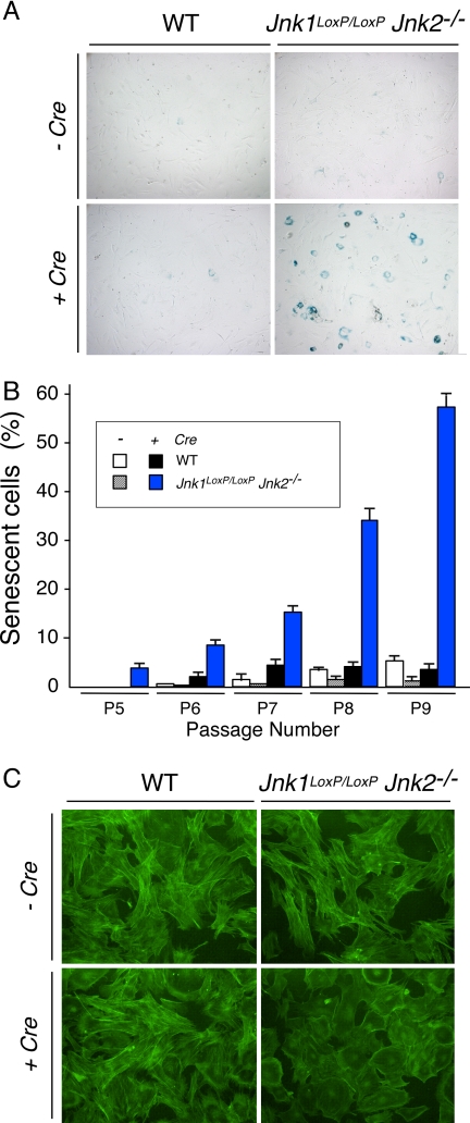
JNK deficiency causes senescence. (A) WT and Jnk1LoxP/LoxP Jnk2−/− MEF were transduced by using a Cre retroviral expression vector and stained for a senescence marker (acidic β-gal) using X-Gal. A culture stained at 3T3 passage 9 postinfection is illustrated. (B) The percentage of cells that stained with X-Gal was examined at passages 5, 6, 7, 8, and 9 postinfection was quantitated. The data shown represent the mean ± SD of three independent experiments. (C) MEF at 3T3 passage 9 postinfection were stained with phalloidin (green) and examined by fluorescence microscopy. (Magnification: ×100.)
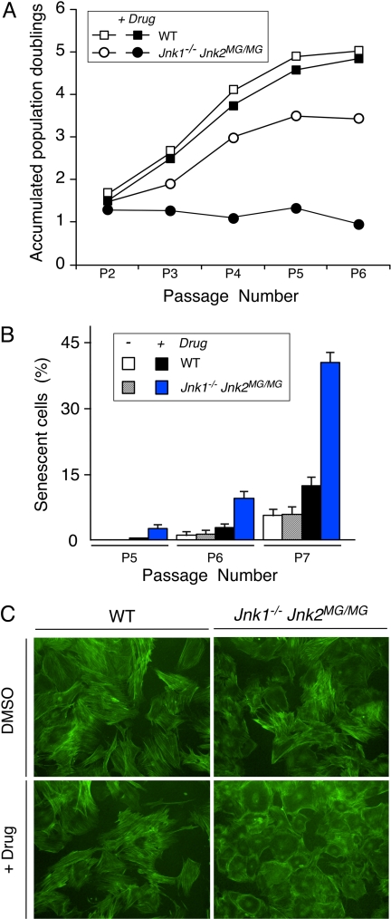
Inhibition of the protein kinase activity of JNK causes senescence. (A) The growth of WT and Jnk1−/− Jnk2MG/MG MEF was examined by using a 3T3 assay. The data are presented as the accumulated population doublings during six passages in culture. The effect of addition of solvent (DMSO) or drug (1 μM 1NM-PP1) was investigated. (B) WT and Jnk1−/− Jnk2MG/MG MEF were stained for a senescence marker (acidic β-gal) by using X-Gal. The percentage of cells that stained with X-Gal was examined at 3T3 passages 5, 6, and 7 and quantitated. The data shown represent the mean ± SD of three independent experiments. (C) MEF at 3T3 passage 7 were stained with phalloidin (green) and examined by fluorescence microscopy. (Magnification: ×100.)
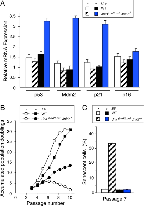
JNK deficiency causes p53-dependent senescence. (A) WT and Jnk1LoxP/LoxP Jnk2−/− MEF were transduced by using a Cre retroviral expression vector. Total RNA was isolated from MEF at 3T3 passage 7 postinfection and the amount of p53, Mdm2, p21, p16, and GAPDH mRNA was measured by quantitative RT-PCR (Taqman assays). The data are presented as the normalized ratio of [p53, Mdm2, p21, or p16 mRNA]/[GAPDH mRNA]. (B) The growth of WT and Jnk1LoxP/LoxP Jnk2−/− MEF (transduced with a retroviral Cre vector) was examined by using a 3T3 assay. The effect of expression of the HPV16 E6 protein is presented. The data are presented as the accumulated population doublings during 10 passages in culture postinfection. (C) WT and Jnk1LoxP/LoxP Jnk2−/− MEF (transduced with a retroviral Cre vector) were stained for a senescence marker (acidic β-gal) by using X-Gal. The effect of expression of the HPV16 E6 protein is presented. The percentage of cells that stained with X-Gal was examined at 3T3 passage 7 postinfection and quantitated. The data shown represent the mean ± SD of three independent experiments.
References
-
- Davis RJ. Cell. 2000;103:239–252. - PubMed
-
- Jochum W, Passegue E, Wagner EF. Oncogene. 2001;20:2401–2412. - PubMed
-
- Tournier C, Hess P, Yang DD, Xu J, Turner TK, Nimnual A, Bar-Sagi D, Jones SN, Flavell RA, Davis RJ. Science. 2000;288:870–874. - PubMed
-
- Manning AM, Davis RJ. Nat Rev Drug Discov. 2003;2:554–565. - PubMed
Publication types
MeSH terms
Substances
LinkOut - more resources
Full Text Sources
Molecular Biology Databases
Research Materials
Miscellaneous
