. 2007 Oct 2;104(40):15619-24.
doi: 10.1073/pnas.0702576104. Epub 2007 Sep 24.

Physical plasticity of the nucleus in stem cell differentiation

Affiliations

Physical plasticity of the nucleus in stem cell differentiation

J David Pajerowski et al. Proc Natl Acad Sci U S A. .

Abstract

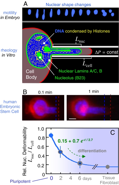
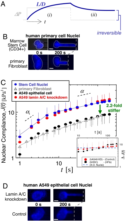
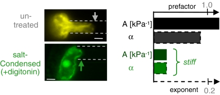
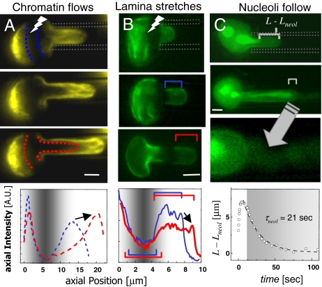
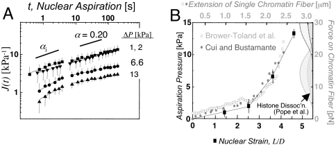
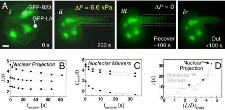
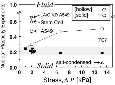
Cell differentiation in embryogenesis involves extensive changes in gene expression structural reorganization within the nucleus, including chromatin condensation and nucleoprotein immobilization. We hypothesized that nuclei in naive stem cells would therefore prove to be physically plastic and also more pliable than nuclei in differentiated cells. Micromanipulation methods indeed show that nuclei in human embryonic stem cells are highly deformable and stiffen 6-fold through terminal differentiation, and that nuclei in human adult stem cells possess an intermediate stiffness and deform irreversibly. Because the nucleo-skeletal component Lamin A/C is not expressed in either type of stem cell, we knocked down Lamin A/C in human epithelial cells and measured a deformability similar to that of adult hematopoietic stem cells. Rheologically, lamin-deficient states prove to be the most fluid-like, especially within the first approximately 10 sec of deformation. Nuclear distortions that persist longer than this are irreversible, and fluorescence-imaged microdeformation with photobleaching confirms that chromatin indeed flows, distends, and reorganizes while the lamina stretches. The rheological character of the nucleus is thus set largely by nucleoplasm/chromatin, whereas the extent of deformation is modulated by the lamina.


Conflict of interest statement

The authors declare no conflict of interest.

Figures

Fig. 1.
Fig. 1.
Nuclei of human stem cells are more deformable than nuclei of differentiated cells. (A Upper) Neural progenitor cell nuclei (12) show large deformations during in vivo migration (SI Fig. 8). Micropipette aspiration mimics such distortions. (B) Human ESCs were aspirated after differentiation in culture, and the ratio of nuclear extension to cytoplasmic extension was measured as Lnuc/Lcell. A day-0 pluripotent ESC is shown with fluorescent dyes labeling nuclear DNA (blue) and the cell membrane (red). (Scale bar: 3 μm.) Dead cells were excluded by counterstaining with propidium iodide. (C) As differentiation progresses, ESC nuclei stiffen nearly 6-fold relative to cytoplasm, and the decrease in relative compliance fits an exponential decay. Differentiated cells such as embryonic fibroblasts also have a nucleus that is stiffer than the cytoplasm. Aspiration of at least three cells and two to three time points per cell were analyzed for each “day” (average ± SD).
Fig. 2.
Fig. 2.
Marrow-derived human HSCs have more compliant nuclei than primary human fibroblasts, which may correspond to a lack of Lamin A/C. (A) Constant pressure aspiration into a micropipette (diameter, D) with creep compliance response for relative aspirated length L/D (i). Release of ΔP and incomplete recovery of L/D (ii). (B–D) After 200 sec of aspiration, HSC nuclei (blue) have deformed 2.2-fold more than fibroblast nuclei (gray), indicating that the latter are stiffer. A549 cells (black) are made equally compliant by knockdown of Lamin A/C (red) (average ± SD; n = 15–20 nuclei). (Scale bars: 3 μm.)
Fig. 3.
Fig. 3.
Divalent salts condense chromatin and stiffen the nucleus. Compared with untreated nuclei, quantification reveals decreases in both the creep prefactor, A, and the creep exponent, α, due to cation-induced condensation. (Scale bar: 3 μm.)
Fig. 4.
Fig. 4.
Distinctive mechanics of nuclear components. (A) FRAP of GFP-H2B chromatin (pseudocolored yellow) reveals chromatin flow within the pipette. Chromatin compaction at the tip is quantified in the axial intensity profiles, and the arrow indicates an increase in intensity with compaction. (B) The lamina (green) is stably stretched into the pipette, and the arrow indicates a decrease in intensity with dilation. (C) Nucleoli slowly follow chromatin toward the nuclear tip, as GFP permeates the intact envelope (arrow into magnified view in bottom panel). (Scale bar: 3 μm.)
Fig. 5.
Fig. 5.
Scale-free creep of nuclei, and nonlinear stress-strain mapping onto chromatin fiber data. (A) Nuclear creep in TC7 cells as a function of pressure (average ± SD; n = 10–15 nuclei). At large ΔP, J shows a crossover at τplastic ≅ 8–10 sec. (B) Overlay of nuclear aspiration results and single chromatin fiber extension data (13, 14). The data from Brower-Toland et al. is normalized to the initial length of the Cui and Bustamante fiber, and stretched by a factor of 1.75. The histograms on the right indicate force-dependent frequency of histone dissociation (15).
Fig. 6.
Fig. 6.
In situ irreversibility of stretched nuclei. (A) The undeformed nucleus (i) is aspirated for ≈200 sec (ii). Recovery within the micropipette for ≈100 sec (iii) is followed by ejection from the pipette (iv). (Scale bar: 3 μm.) Plasticity is evident in the persistently deformed shapes of nuclei and nucleoli, and the time required for even partial recovery increases for both (B and C) at large deformations. (D) Extrapolation of the data to the small strain limit yields τrecover ≅ 8 sec.
Fig. 7.
Fig. 7.
Modulation of nuclear plasticity. Initial deformation response at small stress is more fluid for stem cells and Lamin A/C knock down cells, but differentiated nuclei also display increasingly fluid responses at larger stress. At longer times, all nuclei are more solid-like, with a weak power law exponent of ≈0.2 that is typical of macromolecular networks.

References

    1. Blau H, Pavlath G, Hardeman E, Chiu C, Silberstein L, Webster S, Miller S, Webster C. Science. 1985;230:758–766. - PubMed
    1. Slack JMW, Tosh D. Curr Opin Genet Dev. 2001;11:581–586. - PubMed
    1. Szutorisz H, Dillon N. BioEssays. 2005;27:1286–1293. - PubMed
    1. Labrador M, Corces VG. Cell. 2002;111:151–154. - PubMed
    1. West AG, Fraser P. Hum Mol Genet. 2005;14:R101–R111. - PubMed

Publication types

MeSH terms

Substances