. 2007 Oct 2;104(40):15817-22.
doi: 10.1073/pnas.0706942104. Epub 2007 Sep 24.

Epicutaneous antigen exposure induces a Th17 response that drives airway inflammation after inhalation challenge

Affiliations

Epicutaneous antigen exposure induces a Th17 response that drives airway inflammation after inhalation challenge

Rui He et al. Proc Natl Acad Sci U S A. .

Abstract

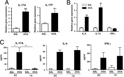
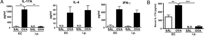
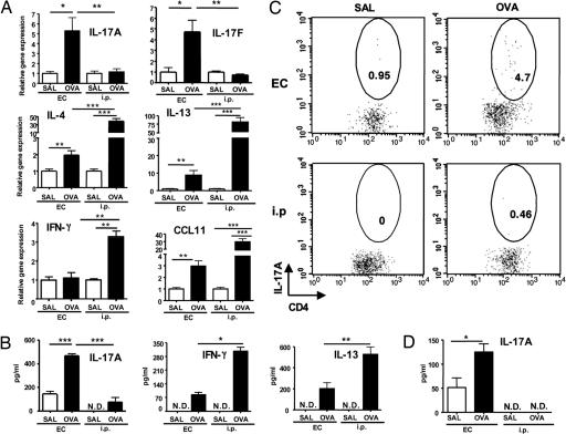
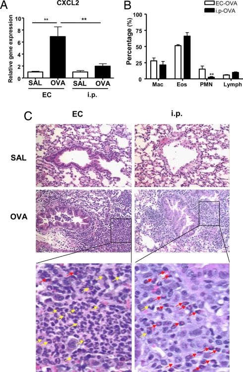
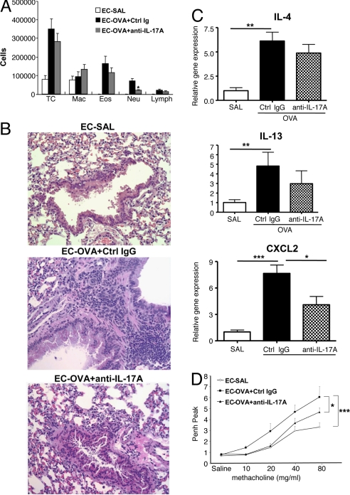
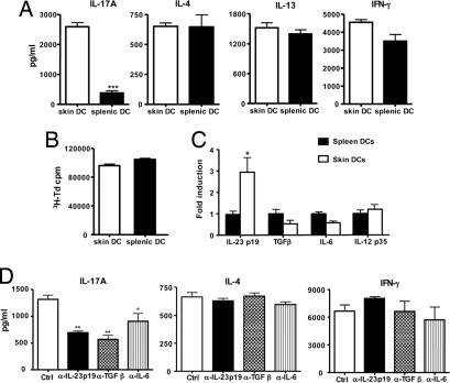
IL-17 has been implicated in a number of inflammatory diseases, but the conditions of antigen exposure that drive the generation of Th17 responses have not been well defined. Epicutaneous (EC) immunization of mice with ovalbumin (OVA), which causes allergic skin inflammation with many characteristics of the skin lesions of atopic dermatitis, was found to also drive IL-17 expression in the skin. EC, but not i.p., immunization of mice with OVA drove the generation of IL-17-producing T cells in draining lymph nodes and spleen and increased serum IL-17 levels. OVA inhalation by EC-sensitized mice induced IL-17 and CXCL2 expression and neutrophil influx in the lung along with bronchial hyperreactivity, which were reversed by IL-17 blockade. Dendritic cells trafficking from skin to lymph nodes expressed more IL-23 and induced more IL-17 secretion by naïve T cells than splenic dendritic cells. This was inhibited by neutralizing IL-23 in vitro and by intradermal injection of anti-TGFbeta neutralizing antibody in vivo. Our findings suggest that initial cutaneous exposure to antigens in patients with atopic dermatitis may selectively induce the production of IL-17, which, in turn, drives inflammation of their airways.


Conflict of interest statement

The authors declare no conflict of interest.

Figures

Fig. 1.
Fig. 1.
Expression of IL-17 in EC-sensitized skin and draining LN. (A and B) Expression of mRNA for IL-17A and IL-17F (A) and of mRNA for IL-4, IL-13, and IFN-γ (B) in sensitized skin with OVA or saline controls (n = 5–7 per group). Values for saline-sensitized sites have been set to 1. (C) IL-17A, IL-4, and IFN-γ secretion by OVA stimulated cells from draining LN. Columns and error bars represent mean± SEM (n = 5–7 per group). *, P < 0.05; **, P < 0.01. N.D., not detectable.
Fig. 2.
Fig. 2.
EC sensitization elicits a systemic Th-17. (A) IL-17A, IL-4, and IFN-γ secretion by OVA-stimulated splenocytes. (B) Serum IL-17A levels in EC versus i.p. sensitized mice. Columns and error bars represent mean± SEM (n = 4–7 per group). *, P < 0.05; **, P < 0.01; ***, P < 0.0001. N.D., not detectable; SAL, saline.
Fig. 3.
Fig. 3.
IL-17 expression in allergen-challenged lungs of EC-sensitized and i.p.-immunized mice. (A) IL-17A, IL-17F, IL-4, IL-13, CCL11, and IFN-γ mRNA expression in lung tissues. Values for lungs of saline sensitized mice have been set to 1. (B) IL-17A, IL-13, and IFN-γ secretion by OVA-stimulated lung cells. (C) Intracellular IL-17A staining of CD4+ cells from OVA-stimulated lung cells. (D) IL-17A levels in bronchoalveolar lavage (BAL) fluid. Columns and error bars represent mean ± SEM (n = 5–6 per group). *, P < 0.05; **, P < 0.01; ***, P < 0.0001. Data in C are representative of two experiments. N.D., not detectable; SAL, saline. Numbers in circled areas indicate the percentage of IL-17+CD4+ cells
Fig. 4.
Fig. 4.
Neutrophil influx in allergen-challenged lungs of EC-sensitized and i.p.-immunized mice. (A) CXCL2 mRNA expression in lung tissues. Values for lungs of saline-sensitized mice have been set to 1. (B) Percentage distribution of cell types in BALF. (C) H&E staining of lung sections from mice EC- and i.p.-sensitized with saline or OVA after OVA-inhalation challenge. Magnification ×200. Further magnification of the black-bordered box shows the predominance of neutrophils (yellow arrows) in lungs from OVA EC-sensitized mice and of eosinophils (red arrows) in lungs from OVA i.p.-sensitized mice. Columns and error bars represent means ± SEM (n = 5–6 per group). *, P < 0.05; **, P < 0.01. SAL, saline.
Fig. 5.
Fig. 5.
Effect of anti-IL-17A on cellular composition of bronchoalveolar lavage fluid, CXCL2 and cytokine expression, and Penh to allergen inhalation in EC-sensitized mice. (A) Cells in bronchoalveolar lavage fluid. (B) Expression of mRNA for CXCL2, IL-4, IL-13, and IFN-γ. Values for lungs of saline-sensitized mice have been set to 1. (C) H&E staining of lung sections examined at magnification ×200 magnification. (D) Peak Penh after OVA-inhalation challenge. Columns and error bars represent means ± SEM (n = 5 per group). *, P < 0.05; **, P < 0.01; ***, P < 0.0001. SAL, saline.
Fig. 6.
Fig. 6.
Selective induction of IL-17A secretion in DO11.1 CD4+ cells by skin-derived DCs. (A) Secretion of IL-17A, IL-4, IL-13, and IFN-γ by DO11.1 CD4+ cells stimulated with OVA323–339 peptide in the presence of skin- or spleen-derived DCs. (B) Proliferation; columns, and error bars represent means of triplicate wells. (C) Q-PCR analysis of mRNA expression of IL-23/p19, TGFβ, IL-6, and IL-12/p35 in spleen- and skin-derived DCs. Values for splenic DCs have been set to 1. (D) Effect of neutralizing antibodies to IL-23p19, TGFβ, and IL-6 on the capacity of skin-derived DCs to induce secretion of IL-17A, IL-4, and IFN-γ by DO11.1 CD4+ stimulated with OVA323–339 peptide. Except for B, columns and error bars represent means ± SEM of three experiments (n = 3); in B, they represent mean ± SEM of triplicates of one experiment representative of three. *, P < 0.05; **, P < 0.01.; ***, P < 0.0001

References

    1. Park H, Li Z, Yang XO, Chang SH, Nurieva R, Wang YH, Wang Y, Hood L, Zhu Z, Tian Q, et al. Nat Immunol. 2005;6:1133–1141. - PMC - PubMed
    1. Harrington LE, Hatton RD, Mangan PR, Turner H, Murphy TL, Murphy KM, Weaver CT. Nat Immunol. 2005;6:1123–1132. - PubMed
    1. Kolls JK, Kanaly ST, Ramsay AJ. Am J Respir Cell Mol Biol. 2003;28:9–11. - PubMed
    1. Veldhoen M, Hocking RJ, Atkins CJ, Locksley RM, Stockinger B. Immunity. 2006;24:179–189. - PubMed
    1. Veldhoen M, Stockinger B. Trends Immunol. 2006;27:358–361. - PubMed

Publication types

LinkOut - more resources