. 2007 Oct;16(10):2093-107.
doi: 10.1110/ps.073011407.

Solution structure and proposed domain domain recognition interface of an acyl carrier protein domain from a modular polyketide synthase

Affiliations

Solution structure and proposed domain domain recognition interface of an acyl carrier protein domain from a modular polyketide synthase

Viktor Y Alekseyev et al. Protein Sci. 2007 Oct.

Abstract

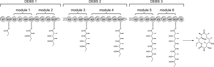
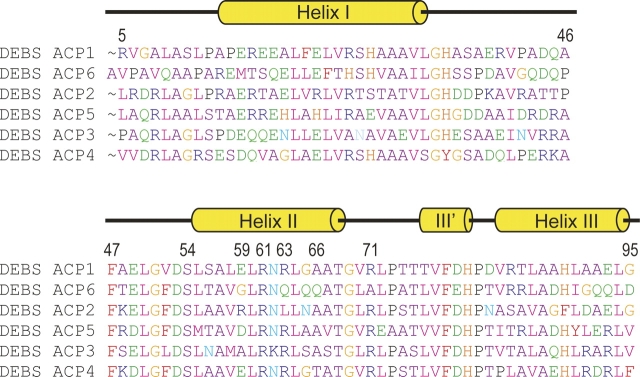
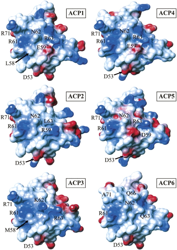
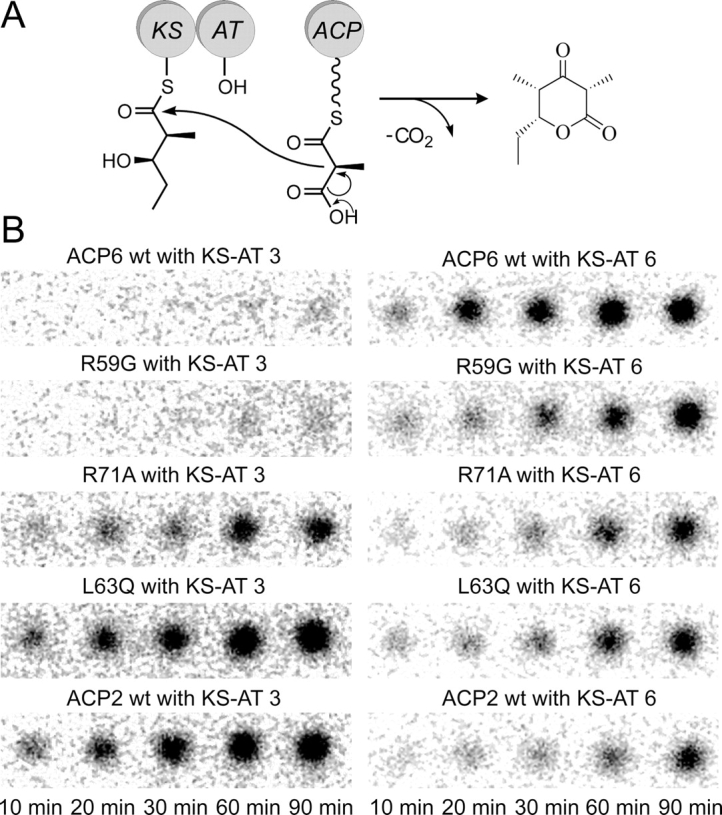
Polyketides are a medicinally important class of natural products. The architecture of modular polyketide synthases (PKSs), composed of multiple covalently linked domains grouped into modules, provides an attractive framework for engineering novel polyketide-producing assemblies. However, impaired domain-domain interactions can compromise the efficiency of engineered polyketide biosynthesis. To facilitate the study of these domain-domain interactions, we have used nuclear magnetic resonance (NMR) spectroscopy to determine the first solution structure of an acyl carrier protein (ACP) domain from a modular PKS, 6-deoxyerythronolide B synthase (DEBS). The tertiary fold of this 10-kD domain is a three-helical bundle; an additional short helix in the second loop also contributes to the core helical packing. Superposition of residues 14-94 of the ensemble on the mean structure yields an average atomic RMSD of 0.64 +/- 0.09 Angstrom for the backbone atoms (1.21 +/- 0.13 Angstrom for all non-hydrogen atoms). The three major helices superimpose with a backbone RMSD of 0.48 +/- 0.10 Angstrom (0.99 +/- 0.11 Angstrom for non-hydrogen atoms). Based on this solution structure, homology models were constructed for five other DEBS ACP domains. Comparison of their steric and electrostatic surfaces at the putative interaction interface (centered on helix II) suggests a model for protein-protein recognition of ACP domains, consistent with the previously observed specificity. Site-directed mutagenesis experiments indicate that two of the identified residues influence the specificity of ACP recognition.


Figures

Figure 1.
Figure 1.
Schematic diagram of the modular organization of 6-deoxyerythronolide B synthase (DEBS). Three polypeptides (DEBS1, DEBS2, and DEBS3) each contain two modules that are, in turn, composed of distinct catalytic domains. KS indicates ketosynthase; AT, acyl transferase; KR, ketoreductase; ACP, acyl carrier protein; DH, dehydratase; ER, enoyl reductase; and TE, thioesterase. DEBS1 also contains a loading didomain, consisting of an AT and an ACP. KR0 denotes an inactive KR domain. Inter-protein linker (docking) domains are shown as matching tabs.
Figure 2.
Figure 2.
Assigned two-dimensional 1H-15N HSQC spectrum of apo-ACP2(Ø) from DEBS. Backbone amide resonances are labeled with the one-letter amino acid code and residue number. Pairs of peaks from asparagine side-chain amide groups are connected by horizontal lines and labeled.
Figure 3.
Figure 3.
Summary of sequential and medium-range NOE patterns for DEBS ACP2(Ø). The relative intensities of sequential NOEs are indicated by the heights of the connecting boxes. Medium-range NOEs observed between residue pairs are indicated by horizontal lines. Helical regions are indicated above the amino acid sequence.
Figure 4.
Figure 4.
NMR solution structure of DEBS ACP2(Ø). (A) Superposition of residues 14–95 for the 30 conformers from torsion angle dynamics calculation. (B) Ribbon diagram of the minimized mean structure of ACP2(Ø). (C) Electrostatic potential surface of ACP2(Ø). Positively charged areas are colored in blue, negatively charged areas are in red, and uncharged areas are in gray. Secondary structure elements are labeled as follows: helices—HI, HII′, HII, HIII′, HIII; loops—LI, LII.
Figure 5.
Figure 5.
Sequence alignment of the DEBS ACP domains. The protein sequences were obtained from the PKSDB (a Database of Modular Polyketide Synthases, www.nii.res.in/pksdb.html). Secondary structure elements from ACP2 structure are shown above the sequences. Several residues discussed in the text are numbered, including the conserved serine (Ser54) that serves as the phosphopantetheine attachment site.
Figure 6.
Figure 6.
Electrostatic potential surface diagrams for six DEBS ACP domains. Positively charged residues are colored blue and negatively charged residues are colored red. Residue numbers for the labeled residues correspond to ACP2 numbering.
Figure 7.
Figure 7.
Triketide lactone formation assay. (A) Chain elongation reaction catalyzed by a KS–AT didomain interacting with a methylmalonyl-ACP. The KS domain is preacylated with the electrophilic diketide substrate (NDK). (B) TLC phosphorimaging results, showing the amount of 14C-labeled triketide lactone product in each reaction at the indicated time points.
Figure 8.
Figure 8.
A phylogram illustrating the clustering of ACP domains into subfamilies. The first 10 ACPs are from modular polyketide synthases: either erythromycin PKS (labeled DebsACP) or rifamycin PKS (labeled RifACP). The ACP domains from fatty acid synthases (E. coli ACP and B. subtilis ACP) form a distinct subgroup. In turn, the actinorhodin (act), frenolicin (fren), and oxytetracycline (otc) ACPs, which originate from type II (aromatic) polyketide synthases, also form a subfamily distinct both from the ACP domains of the fatty acid synthase origin and from the ACPs that belong to modular polyketide synthases. The sequences were aligned using PileUp program (GCG SeqWeb software package, Accelrys Inc.) and the phylogram was produced using the CLUSTAL W server at the European Bioinformatics Institute (www.ebi.ac.uk/clustalw/).

References

    1. Broadhurst R.W., Nietlispach, D., Wheatcroft, M.P., Leadlay, P.F., and Weissman, K.J. 2003. The structure of docking domains in modular polyketide synthases. Chem. Biol. 10: 723–731. - PubMed
    1. Cane D.E., Walsh, C.T., and Khosla, C. 1998. Harnessing the biosynthetic code: Combinations, permutations, and mutations. Science 282: 63–68. - PubMed
    1. Cortes J., Haydock, S.F., Roberts, G.A., Bevitt, D.J., and Leadlay, P.F. 1990. An unusually large multifunctional polypeptide in the erythromycin-producing polyketide synthase of Saccharopolyspora erythraea . Nature 348: 176–178. - PubMed
    1. Crump M.P., Crosby, J., Dempsey, C.E., Parkinson, J.A., Murray, M., Hopwood, D.A., and Simpson, T.J. 1997. Solution structure of the actinorhodin polyketide synthase acyl carrier protein from Streptomyces coelicolor A3(2) . Biochemistry 36: 6000–6008. - PubMed
    1. Donadio S. and Katz, L. 1992. Organization of the enzymatic domains in the multifunctional polyketide synthase involved in erythromycin formation in Saccharopolyspora erythraea . Gene 111: 51–60. - PubMed

Publication types

MeSH terms

LinkOut - more resources