

doi: 10.1110/ps.073018907.
WrbA bridges bacterial flavodoxins and eukaryotic NAD(P)H:quinone oxidoreductases
Affiliations
- PMID: 17893367
- PMCID: PMC2204128
- DOI: 10.1110/ps.073018907
Item in Clipboard
WrbA bridges bacterial flavodoxins and eukaryotic NAD(P)H:quinone oxidoreductases
Protein Sci.
2007 Oct.
Abstract
The crystal structure of the flavodoxin-like protein WrbA with oxidized FMN bound reveals a close relationship to mammalian NAD(P)H:quinone oxidoreductase, Nqo1. Structural comparison of WrbA, flavodoxin, and Nqo1 indicates how the twisted open-sheet fold of flavodoxins is elaborated to form multimers that extend catalytic function from one-electron transfer between protein partners using FMN to two-electron reduction of xenobiotics using FAD. The structure suggests a novel physiological role for WrbA and Nqo1.
Figures
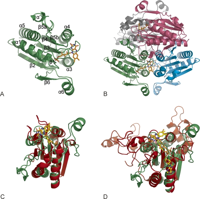
WrbA bridges flavodoxins and oxidoreductases. (A) Overall fold. WrbA monomer, ribbon; FMN, skeletal (orange carbon atoms and atomic colors). Secondary structure numbering follows the convention introduced for Nqo1 (Li et al. 1995). Crystallization and diffraction analysis are described by Wolfová et al. (2007). (B) WrbA tetramer. View along the diagonal between crystallographic axes a and b, with each subunit a unique color. (C) Overlay with flavodoxin. Least-squares 3D alignment of WrbA monomer (rotated 90° from panel A) with short-chain flavodoxin (PDB ID 1J8Q; protein, red; FMN, yellow carbons and atomic colors). (D) Overlay with Nqo1. Mammalian Nqo1 monomer (PDB ID 1QRD; protein, red; FAD, yellow carbons and atomic colors). C-terminal subdomain at upper left.
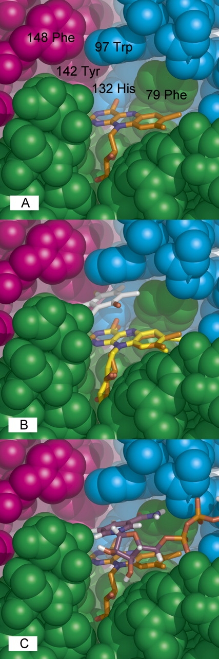
WrbA active site. (A) Key residues. Space-filling representation with residues numbered and chain colors as in Figure 1B. (B) Quinone binding. The WrbA active site was overlaid with that of 1QRD with bound duroquinone (Li et al. 1995) by superposition of the common flavin atoms from isoalloxazine through phosphate. (C) NADH binding. The program FlexX (Rarey et al. 1996) was used to dock NADH into the WrbA tetramer (nicotinamide and adenine riboside carbons, purple; pyrophosphate, orange; other colors atomic).
References
-
- Asher G., Dym, O., Tsvetkov, P., Adler, J., and Shaul, Y. 2006. The crystal structure of NAD(P)H:quinone oxidoreductase 1 in complex with its potent inhibitor dicoumarol. Biochemistry 45: 6372–6378. - PubMed
-
- Beyer R.E., Segura-Aguilar, J., DiBernardo, S., Cavazzoni, M., Fato, R., Fiorentini, D., Galli, M.C., Setti, M., Landi, L., and Lenaz, G. 1996. The role of DT-diaphorase in the maintenance of the reduced antioxidant form of coenzyme-Q in membrane systems. Proc. Natl. Acad. Sci. 93: 2528–2532. - PMC - PubMed
-
- Cadenas E. 1995. Antioxidant and prooxidant functions of DT-diaphorase in quinone metabolism. Biochem. Pharmacol. 49: 127–140. - PubMed
-
- Ernster L. 1987. DT-Diaphorase: A historical review. Chem. Scr. 27A: 1–13.
-
- Georgellis D., Kwon, O., and Lin, E.C. 2001. Quinones as the redox signal for the arc two-component system of bacteria. Science 292: 2314–2316. - PubMed
Publication types
MeSH terms
Substances
LinkOut - more resources
Full Text Sources
Molecular Biology Databases
Miscellaneous
