

Mapping functional connectivity in patients with brain lesions
- PMID: 17894381
- PMCID: PMC3646715
- DOI: 10.1002/ana.21224
Mapping functional connectivity in patients with brain lesions
Abstract
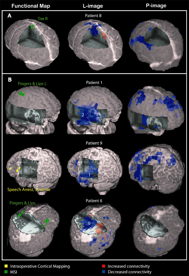
Objective: The spatial distribution of functional connectivity between brain areas and the disturbance introduced by focal brain lesions are poorly understood. Based on the rationale that damaged brain tissue is disconnected from the physiological interactions among healthy areas, this study aimed to map the functionality of brain areas according to their connectivity with other areas.
Methods: Magnetoencephalography recordings of spontaneous cortical activity during resting state were obtained from 15 consecutive patients with focal brain lesions and from 14 healthy control subjects. Neural activity in the brain was estimated using an adaptive spatial filtering technique. The mean imaginary coherence between brain voxels was then calculated as an index of functional connectivity.
Results: Imaginary coherence was greatest in the alpha frequency range corresponding to the human cortical idling rhythm. In healthy subjects, functionally critical brain areas such as the somatosensory and language cortices had the highest alpha coherence. When compared with healthy control subjects, all lesion patients had diffuse or scattered brain areas with decreased alpha coherence. Patients with lesion-induced neurological deficits displayed decreased connectivity estimates in the corresponding brain area compared with intact contralateral regions. In tumor patients without preoperative neurological deficits, brain areas showing decreased coherence could be surgically resected without the occurrence of postoperative deficits.
Interpretation: Resting state coherence measured with magnetoencephalography is capable of mapping the functional connectivity of the brain, and can therefore offer valuable information for use in planning resective surgeries in patients with brain lesions, as well as investigations into structural-functional relationships in healthy subjects.
Figures


References
-
- Alberstone CD, Skirboll SL, Benzel EC, et al. Magnetic source imaging and brain surgery: presurgical and intraoperative planning in 26 patients. J Neurosurg. 2000;92:79–90. - PubMed
-
- Bartolomei F, Bosma I, Klein M, et al. Disturbed functional connectivity in brain tumour patients: evaluation by graph analysis of synchronization matrices. Clin Neurophysiol. 2006a;117:2039–2049. - PubMed
-
- Bartolomei F, Bosma I, Klein M, et al. How do brain tumors alter functional connectivity? A magnetoencephalography study. Ann Neurol. 2006b;59:128–138. - PubMed
-
- Berger MS, Kincaid J, Ojemann GA, Lettich E. Brain mapping techniques to maximize resection, safety, and seizure control in children with brain tumors. Neurosurgery. 1989;25:786–792. - PubMed
-
- Fandino J, Kollias SS, Wieser HG, Valavanis A, Yonekawa Y. Intraoperative validation of functional magnetic resonance imaging and cortical reorganization patterns in patients with brain tumors involving the primary motor cortex. J Neurosurg. 1999;91:238–250. - PubMed
Publication types
MeSH terms
Grants and funding
LinkOut - more resources
Full Text Sources
Medical
