. 2007 Sep 25:4:9.
doi: 10.1186/1743-8977-4-9.

Translocation of particles and inflammatory responses after exposure to fine particles and nanoparticles in an epithelial airway model

Affiliations

Translocation of particles and inflammatory responses after exposure to fine particles and nanoparticles in an epithelial airway model

Barbara Rothen-Rutishauser et al. Part Fibre Toxicol. .

Abstract

Background: Experimental studies provide evidence that inhaled nanoparticles may translocate over the airspace epithelium and cause increased cellular inflammation. Little is known, however, about the dependence of particle size or material on translocation characteristics, inflammatory response and intracellular localization.

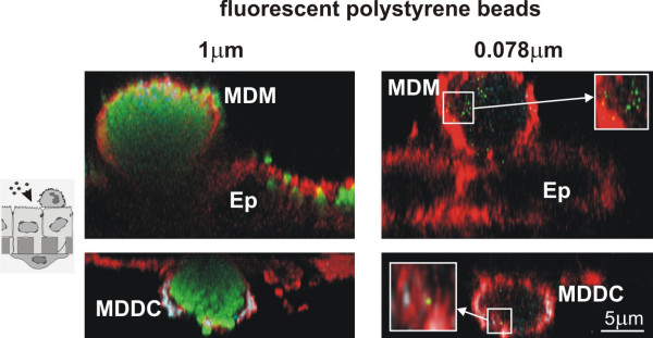
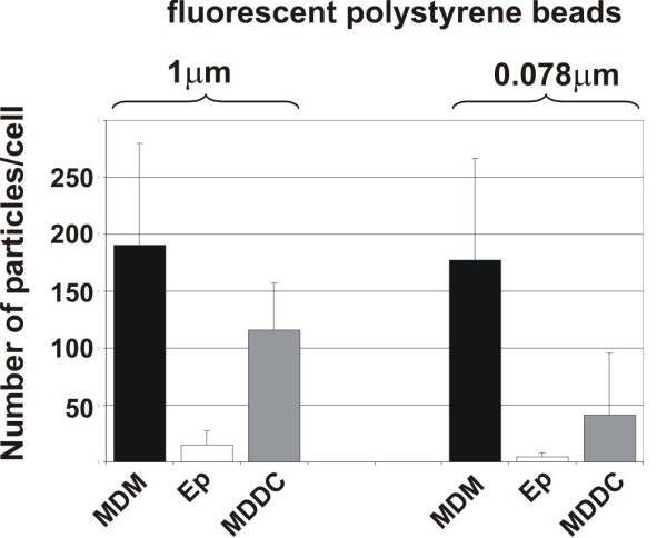
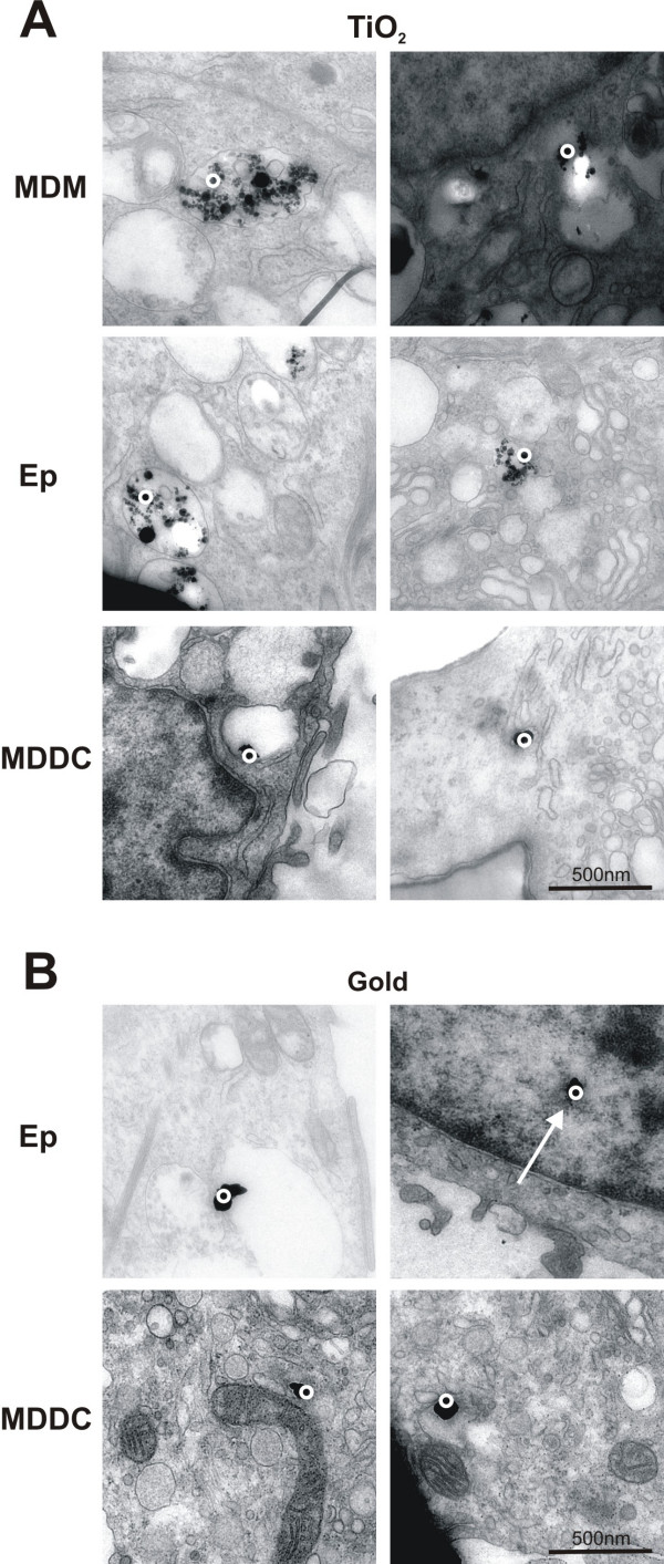
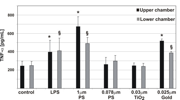
Results: Using a triple cell co-culture model of the human airway wall composed of epithelial cells, macrophages and dendritic cells we quantified the entering of fine (1 mum) and nano-sized (0.078 mum) polystyrene particles by laser scanning microscopy. The number distribution of particles within the cell types was significantly different between fine and nano-sized particles suggesting different translocation characteristics. Analysis of the intracellular localization of gold (0.025 mum) and titanium dioxide (0.02-0.03 mum) nanoparticles by energy filtering transmission electron microscopy showed differences in intracellular localization depending on particle composition. Titanium dioxide nanoparticles were detected as single particles without membranes as well as in membrane-bound agglomerations. Gold nanoparticles were found inside the cells as free particles only. The potential of the different particle types (different sizes and different materials) to induce a cellular response was determined by measurements of the tumour necrosis factor-alpha in the supernatants. We measured a 2-3 fold increase of tumour necrosis factor-alpha in the supernatants after applying 1 mum polystyrene particles, gold nanoparticles, but not with polystyrene and titanium dioxide nanoparticles.

Conclusion: Quantitative laser scanning microscopy provided evidence that the translocation and entering characteristics of particles are size-dependent. Energy filtering transmission electron microscopy showed that the intracellular localization of nanoparticles depends on the particle material. Both particle size and material affect the cellular responses to particle exposure as measured by the generation of tumour necrosis factor-alpha.


Figures

Figure 1
Figure 1
Intracellular particle localisation in tripe cell co-cultures visualised by LSM. Cells at the upper side of the insert (upper row) were stained for CD14 (MDM, turquoise) and F-Actin (all cells, red); cells at the lower side (lower row) for CD86 (MDDC, turquoise) and F-Actin (all cells, red). Fluorescently labelled polystyrene particles (1 μm, and 0.078 μm) are shown in green. MDM and MDDC were filled with particles, considerably fewer particles were found in epithelial cells (Ep). All images represent xz-projections.
Figure 2
Figure 2
Quantification of particles inside individual cells. Intracellular particle numbers were analysed in cultures exposed to 1 μm, and 0.078 μm polystyrene particles with the software Diacount. Epithelial cells (Ep). Number of intracellular particles was counted in individual cells. Data are expressed as the mean of 3–4 experiments (10–14 cells scanned per experiment by LSM).
Figure 3
Figure 3
EELS images of cells containing TiO2 and silver enhanced gold particles. TiO2 particles (A) were found inside all cell types, i.e. MDM, epithelial cells, and MDDC, as aggregates in vesicles (A, left panel), and as single particles or as small aggregates free in the cytoplasm (A, right panel). Silver enhanced gold particles (B) were found in all three cell types as single particles or as small aggregates only free in the cytoplasm and even in the nucleus (B, arrow). The circles mark the region where the element analysis was performed.
Figure 4
Figure 4
TNF-α release in triple cell co-cultures upon particle incubation. TNF-α levels in the supernatants (upper chamber, lower chamber) were measured by ELISA. TNF-α release in cells exposed to LPS, 1 μm, and 0.078 μm polystyrene particles, TiO2, and gold NP. Values are means ± SD of 3 experiments. * indicates a statistical difference to the levels in the supernatants in the control of the upper chamber, § indicates a statistical difference to the levels in the supernatants in the control of the lower chamber.

References

    1. Mazzola L. Commercializing nanotechnology. Nat Biotechnol. 2003;21:1137–1143. doi: 10.1038/nbt1003-1137. - DOI - PubMed
    1. Paull R, Wolfe J, Hebert P, Sinkula M. Investing in nanotechnology. Nat Biotechnol. 2003;21:1144–1147. doi: 10.1038/nbt1003-1144. - DOI - PubMed
    1. Gwinn MR, Vallyathan V. Nanoparticles: Health Effects-Pros and Cons. Environ Health Perspect. 2006;114:1818–1825. - PMC - PubMed
    1. Moghimi SM, Hunter AC, Murray JC. Nanomedicine: current status and future prospects. FASEB J. 2005;19:311–330. doi: 10.1096/fj.04-2747rev. - DOI - PubMed
    1. Maynard A, Michelson E. The Nanotechnology Consumer Products Inventory. 2005. http://www.nanotechproject.org/index.php?id=44