

A model for the role of integrins in flow induced mechanotransduction in osteocytes
- PMID: 17895377
- PMCID: PMC2000405
- DOI: 10.1073/pnas.0707246104
A model for the role of integrins in flow induced mechanotransduction in osteocytes
Abstract
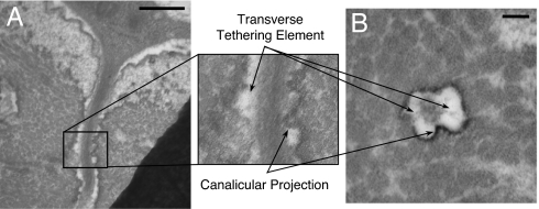
A fundamental paradox in bone mechanobiology is that tissue-level strains caused by human locomotion are too small to initiate intracellular signaling in osteocytes. A cellular-level strain-amplification model previously has been proposed to explain this paradox. However, the molecular mechanism for initiating signaling has eluded detection because none of the molecules in this previously proposed model are known mediators of intracellular signaling. In this paper, we explore a paradigm and quantitative model for the initiation of intracellular signaling, namely that the processes are attached directly at discrete locations along the canalicular wall by beta(3) integrins at the apex of infrequent, previously unrecognized canalicular projections. Unique rapid fixation techniques have identified these projections and have shown them to be consistent with other studies suggesting that the adhesion molecules are alpha(v)beta(3) integrins. Our theoretical model predicts that the tensile forces acting on the integrins are <15 pN and thus provide stable attachment for the range of physiological loadings. The model also predicts that axial strains caused by the sliding of actin microfilaments about the fixed integrin attachments are an order of magnitude larger than the radial strains in the previously proposed strain-amplification theory and two orders of magnitude greater than whole-tissue strains. In vitro experiments indicated that membrane strains of this order are large enough to open stretch-activated cation channels.
Conflict of interest statement
The authors declare no conflict of interest.
Figures


References
-
- Rubin CT, Lanyon LE. J Bone Joint Surg Am. 1984;66:397–402. - PubMed
-
- Fritton SP, McLeod KJ, Rubin CT. J Biomech. 2000;33:317–325. - PubMed
-
- You J, Yellowley CE, Donahue HJ, Zhang Y, Chen Q, Jacobs CR. J Biomech Eng. 2000;122:387–393. - PubMed
-
- Smalt R, Mitchell FT, Howard RL, Chambers TJ. Am J Physiol. 1997;273:E751–E758. - PubMed
-
- Cowin SC, Moss-Salentijn L, Moss ML. J Biomech Eng. 1991;113:191–197. - PubMed
Publication types
MeSH terms
Substances
Grants and funding
LinkOut - more resources
Full Text Sources
Miscellaneous
