Molecular basis for passive immunotherapy of Alzheimer's disease
- PMID: 17895381
- PMCID: PMC1994138
- DOI: 10.1073/pnas.0705888104
Molecular basis for passive immunotherapy of Alzheimer's disease
Abstract
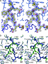
Amyloid aggregates of the amyloid-beta (Abeta) peptide are implicated in the pathology of Alzheimer's disease. Anti-Abeta monoclonal antibodies (mAbs) have been shown to reduce amyloid plaques in vitro and in animal studies. Consequently, passive immunization is being considered for treating Alzheimer's, and anti-Abeta mAbs are now in phase II trials. We report the isolation of two mAbs (PFA1 and PFA2) that recognize Abeta monomers, protofibrils, and fibrils and the structures of their antigen binding fragments (Fabs) in complex with the Abeta(1-8) peptide DAEFRHDS. The immunodominant EFRHD sequence forms salt bridges, hydrogen bonds, and hydrophobic contacts, including interactions with a striking WWDDD motif of the antigen binding fragments. We also show that a similar sequence (AKFRHD) derived from the human protein GRIP1 is able to cross-react with both PFA1 and PFA2 and, when cocrystallized with PFA1, binds in an identical conformation to Abeta(1-8). Because such cross-reactivity has implications for potential side effects of immunotherapy, our structures provide a template for designing derivative mAbs that target Abeta with improved specificity and higher affinity.
Conflict of interest statement
The authors declare no conflict of interest.
Figures


References
-
- Dobson CM. Nature. 2002;418:729–730. - PubMed
-
- Weiner HL, Frenkel D. Nat Rev Immunol. 2006;6:404–416. - PubMed
-
- Gevorkian G, Petrushina I, Manoutcharian K, Ghochikyan A, Acero G, Vasilevko V, Cribbs DH, Agadjanyan MG. J Neuroimmunol. 2004;156:10–20. - PubMed
-
- Solomon B. Curr Alzheimer Res. 2004;1:149–163. - PubMed
Publication types
MeSH terms
Substances
Associated data
- Actions
- Actions
- Actions
- Actions
- Actions
- Actions
Grants and funding
LinkOut - more resources
Full Text Sources
Other Literature Sources
Medical
Molecular Biology Databases

