. 2007 Oct 2;104(40):15699-704.
doi: 10.1073/pnas.0703182104. Epub 2007 Sep 25.

Time-resolved NMR methods resolving ligand-induced RNA folding at atomic resolution

Affiliations

Time-resolved NMR methods resolving ligand-induced RNA folding at atomic resolution

Janina Buck et al. Proc Natl Acad Sci U S A. .

Abstract

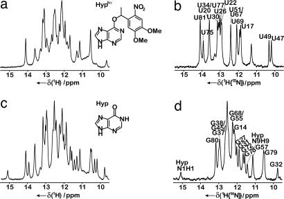
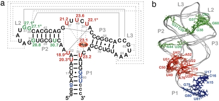
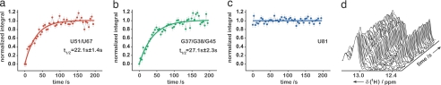
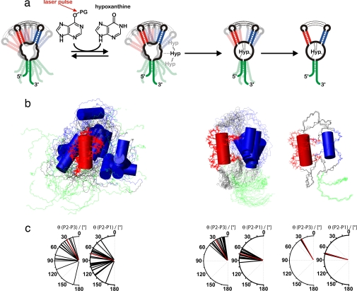
Structural transitions of RNA between alternate conformations with similar stabilities are associated with important aspects of cellular function. Few techniques presently exist that are capable of monitoring such transitions and thereby provide insight into RNA dynamics and function at atomic resolution. Riboswitches are found in the 5'-UTR of mRNA and control gene expression through structural transitions after ligand recognition. A time-resolved NMR strategy was established in conjunction with laser-triggered release of the ligand from a photocaged derivative in situ to monitor the hypoxanthine-induced folding of the guanine-sensing riboswitch aptamer domain of the Bacillus subtilis xpt-pbuX operon at atomic resolution. Combining selective isotope labeling of the RNA with NMR filter techniques resulted in significant spectral resolution and allowed kinetic analysis of the buildup rates for individual nucleotides in real time. Three distinct kinetic steps associated with the ligand-induced folding were delineated. After initial complex encounter the ligand-binding pocket is formed and results in subsequent stabilization of a remote long-range loop-loop interaction. Incorporation of NMR data into experimentally restrained molecular dynamics simulations provided insight into the RNA structural ensembles involved during the conformational transition.


Conflict of interest statement

The authors declare no conflict of interest.

Figures

Fig. 1.
Fig. 1.
Imino proton spectra of GSRapt. (a) Unlabeled RNA before laser pulse. (Inset) Chemical structure of DMNPE-hypoxanthine (Hyp). (b) [15N]uridine-labeled RNA, uridine residues as a result of 1H{15N}-detection after laser pulse with annotated NMR resonance assignment (31) of resolved residues. (c) Unlabeled RNA after laser pulse. (Inset) Chemical chemical structure of hypoxanthine (Hyp). (d) [15N]uridine-labeled RNA, guanosine residues as a result of 1H{14N}-detection after laser pulse with annotated NMR resonance assignment (31) of resolved residues.
Fig. 2.
Fig. 2.
Secondary (a) and tertiary (b) structure of GSRapt with kinetic results. Red, half-life values [t1/2 (s)] in the time range 18.9–23.6 s; green, half-life values in the time range 27.1–30.7 s; blue, signals that remain unaffected during the structural transition; asterisk, overlaid signal (for further information, see text); Hyp, hypoxanthine; labeling of helices P1, P2, and P3 and loop regions L2 and L3, according to Breaker et al. (18); gray solid lines, Watson–Crick base-pairing interactions; gray dashed lines, noncanonical base-pairing interactions [for construct details, see supporting information (SI) Fig. 5].
Fig. 3.
Fig. 3.
Representation of individual signals during time course of reaction. (a–c) Normalized integrals of imino proton signals: (a) red, core region signal U51/U67; (b) green, loop region signal G37/G38/G45; (c) blue, signal U81 that is part of helix P1 as a function of time with monoexponential fit (for signals U51/U67 and G37/G38/G45) and linear fit (for signal U81) (solid line); (d) stack plot of a series of 1H{15N}-NMR spectra as a function of time (imino proton subsection, 12.2–13.4 ppm).
Fig. 4.
Fig. 4.
Structural interpretation of the conformational transition. (a) Schematic illustration of the proposed folding model of GSRapt-RNA on ligand binding based on the experimentally restrained torsion angle MD simulations. The first step is low-affinity binding, the second step is the ligand-binding process, and the third step is helical tightening. (b) Overlaid structures of the three states simulated according to our NMR data, aligned on helix P2 (red). Helices P3 and P1 are blue and green, respectively. (Left) The free form of GSRapt, where only the loop–loop interaction and the canonical form of the three helices are restrained. (Center) The transition state-like form where the core is folded toward the native conformation. (Right) The native structure. (c) Distribution of helix–helix projection angles [°] between helices P2/P3 and P2/P1 as seen in the crystal structure (22) (red) and in the structural ensembles depicted in Fig. 4b (black).

References

    1. Radford SE, Dobson CM, Evans PA. Nature. 1992;358:302–307. - PubMed
    1. Vendruscolo M, Paci E, Dobson CM, Karplus M. Nature. 2001;409:641–645. - PubMed
    1. Salvatella X, Dobson CM, Fersht AR, Vendruscolo M. Proc Natl Acad Sci USA. 2005;102:12389–12394. - PMC - PubMed
    1. Wyatt JR, Puglisi JD, Tinoco I., Jr BioEssays. 1989;11:100–106. - PubMed
    1. Schroeder R, Barta A, Semrad K. Nat Rev Mol Cell Biol. 2004;5:908–919. - PubMed

Publication types