. 2007 Dec;15(12):2154-63.
doi: 10.1038/sj.mt.6300305. Epub 2007 Sep 25.

One-year expression from high-capacity adenoviral vectors in the brains of animals with pre-existing anti-adenoviral immunity: clinical implications

Affiliations

One-year expression from high-capacity adenoviral vectors in the brains of animals with pre-existing anti-adenoviral immunity: clinical implications

Carlos Barcia et al. Mol Ther. 2007 Dec.

Abstract

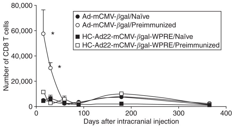
The main challenge of gene therapy is to provide long-term, efficient transgene expression. Long-term transgene expression from first generation adenoviral vectors (Advs) delivered to the central nervous system (CNS) is elicited in animals not previously exposed to adenovirus (Ad). However, upon systemic immunization against Ad, transgene expression from a first generation Adv is abolished. High-capacity Advs (HC-Advs) provide sustained very long-term transgene expression in the brain, even in animals pre-immunized against Ad. In this study, we tested the hypothesis that a HC-Adv in the brain would allow for long-term transgene expression, for up to 1 year, in the brain of mice immunized against Ad prior to delivery of the vector to the striatum. In naïve animals, the expression of beta-galactosidase from Adv or HC-Adv was sustained for 1 year. In animals immunized prior to vector delivery, expression from a first generation Adv was abolished. These results point to a very long-term HC-Adv-mediated transgene expression in the brain, even in animals that had been immunized systemically against Ad before the delivery of HC-Adv into the brain. This study therefore indicates the utility of HC-Adv as a powerful gene therapy vector for chronic neurological disorders, even in patients who had been pre-exposed to Ad prior to gene therapy.


Figures

Figure 1
Figure 1. β-galactosidase (β-gal) expression in neurons and astrocytes in non-immunized brains infected with adenovirus (Ad) (HC-Ad22-mCMV-βgal-WPRE)
Panel (a) illustrates confocal images of immunohistochemistry for β-gal combined with the astrocyte marker glial fibrillary acidic protein (GFAP) (top row) or microtubule-associated protein-2 (MAP-2) (marker for neurons) (second row) of mouse brain sections injected in the striatum with Ad-mCMV-βgal. The top row shows merged images illustrating the co-localization of β-gal and GFAP, thus confirming the expression of transgene in astrocytes (97%). The second row of images shows staining for β-gal combined with MAP-2. In the merged image no co-localization is observed demonstrating that β-gal is not expressed in neurons in mouse brains injected with Ad-mCMV-βgal (0%). Panel (b) shows confocal images of immunofluorescence for β-gal combined with GFAP (marker for astrocytes) (top row) or MAP-2 (marker for neurons) (second row) of sections of mouse brain injected in the striatum with HC-Ad22-mCMV-βgal-WPRE. The top row shows staining for β-gal combined with GFAP. In the merged image note the co-localization of β-gal and GFAP verifying the expression of transgene in astrocytes (70%). The second row of images show staining for β-gal combined with MAP-2. The merged image shows co-localization of β-gal with MAP-2 demonstrating that the transgene is expressed in neurons in the brain of mice injected with HC-Ad22-mCMV-βgal-WPRE (26%). Scale bar: 30 μm. mCMV, murine cytomegalovirus; WPRE, wood chuck hepatitis virus post-transcriptional regulatory element.
Figure 2
Figure 2. Long-term transgene expression following the injection of adenovirus (Ad) into naïve mice, or mice immunized against Ad preceding the delivery of vectors into the brain
Panel (a) shows mouse brain sections injected with Ad-mCMV-βgal immunostained for β-galactosidase (β-gal) in naïve animals (top row), or animals preimmunized against Ad (second row) at different time points. Naïve animals show a robust expression of transgene even 1 year after the intrastriatal injection. However, preimmunized animals show a significant decrease of transgene expression 14 days after injection and finally loss of the transgene expression. Panel (b) shows the expression of β-gal in brain sections of mice injected with High-capacity Ad (HC-Ad) (HC-Ad22-mCMV-βgal-WPRE). Both naïve and preimmunized animals show sustained expression of the transgene even 1 year after vector injection into the brain. Scale bar: 1 mm. (c,d) shows the stereological quantification of β-gal expressing cells in animals injected with Ad-mCMV-βgal or HC-Ad22-mCMV-βgal-WPRE. The quantitative analysis demonstrates that the expression of the transgene is sustained following HC-Ad injection 1 year after the injection even in animals that have been pre-immunized against Adv. c shows the stereological estimation of the total number of β-gal expressing cells in mouse striatum 14, 30, 60, 90, 180, and 365 days after intracranial (IC) injection of Ad-mCMV-βgal in naïve and preimmunized animals. Note that naïve animals show sustained transgene expression that decreases slightly over 1 year. However preimmunized animals show a significant decrease of transgene expression 14 and 30 days after IC injection, and it is almost completely lost at 180 days [(F = 57.08, df = 1, P < 0.001; two way analysis of variance (ANOVA)]. d shows the stereological estimation of the total number of β-gal expressing cells in mouse striatum 14, 30, 60, 90, 180, and 365 days following the IC injection of HC-Ad22-mCMV-βgal-WPRE in naïve and preimmunized animals. Naïve and preimmunized animals illustrate sustained transgene expression over 1 year. There was no significant difference in transgene expression between immunized and non-immunized animals (F = 3.36, df = 1, P = 0.07; two way ANOVA) and expression levels were maintained for 1 year post-vector injection into the brain (Figure 2) (F = 1.2, df = 5, P = 0.33; two way ANOVA). IP, intraperitoneal; mCMV, murine cytomegalovirus; WPRE, wood chuck hepatitis virus post-transcriptional regulatory element.
Figure 3
Figure 3. Anti-adenovirus neutralizing antibody titers in animals injected with Ad into the brain
Titer of neutralizing antibodies against Ad in serum of animals injected either with Ad-mCMV-βgal (a) or HC-Ad22-mCMV-βgal-WPRE (b) in the striatum of animals immunized against Ad (Ad-hCMV-HPRT), a month earlier. Sera were analyzed 14, 30, 60, 90, 180, and 365 days after the intracranial (IC) delivery of vectors. As expected, immunization with Ad-hCMV-HPRT induced a high titer neutralizing antibody response evidenced by approximately 1:200 titer 60 days after the IC injection. Very low titers were found 90 days post injection, which become very low or absent after 180 days. Note that naïve animals (sham-immunized) do not show any neutralizing antibodies against Ad. There were no differences in the serum antibody titers of animals injected into the brain with either vector. Ad, adenovirus; hCMV, human cytomegalovirus; HPRT, hypoxanthine-guanine phosphoribosyltransferase; IP, intraperitoneal; mCMV, murine cytomegalovirus; WPRE, wood chuck hepatitis virus post-transcriptional regulatory element.
Figure 4
Figure 4. Infiltration of CD8+ T cells following the delivery of adenovirus (Ad) into the brain of immunized mice
This graph illustrates the unbiased stereological quantification of CD8+ T cells that infiltrated the striatum of mice preimmunized with Ad-hCMV-HPRT, following the intracranial injection of either Ad-mCMV-βgal or high capacity-Ad (HC-Ad). Preimmunized animals injected intracranially with HC-Ad do not show any statistically significant increase of CD8+ T cells in the striatum. However, preimmunized animals injected intracranially with Ad-mCMV-βgal show a dramatic increase in the number of CD8+ T cells in the striatum 14 and 30 days after the intracranial injection. *P < 0.05 with respect to naïve animals and preimmunized animals injected with HC-Adv. hCMV, human cytomegalovirus; HPRT, hypoxanthine-guanine phosphoribosyltransferase; mCMV, murine cytomegalovirus; WPRE, wood chuck hepatitis virus post-transcriptional regulatory element.
Figure 5
Figure 5. High capacity-adenovirus (HC-Ad) is not able to prime a systemic anti-Ad immune response
(a) shows the serum neutralizing antibody titers against Ad of animals injected with Ad-mCMV-βgal in the striatum and immunized 30 days later, with either Ad (Ad-hCMV-HPRT) or HC-Ad22-mCMV-TKnull-WPRE; brains and sera were analyzed 30 and 60 days after immunization. Note that Ad-hCMV-HPRT immunization is able to generate a high neutralizing antibody response in comparison with the very low or absent titer induced by HC-Ad immunization. Control naïve animals (injected with saline) did not show any neutralizing antibodies against Ad. (b) shows the number of β-galactosidase (β-gal) expressing cells in the striatum of mice injected with Ad-mCMV-βgal in naïve animals (injected with saline; sham immunized), animals immunized with Ad (Ad-hCMV-HPRT), or animals immunized with HC-Ad22-mCMV-TKnull-WPRE. Animals immunized with Ad-hCMV-HPRT show an increase in the serum antibody titer, and a corresponding decrease in the number of β-gal expressing cells. However, animals immunized with HC-Ad show no statistical differences compared to the naïve animals. *P < 0.05 compare to saline. hCMV, human cytomegalovirus; HPRT, hypoxanthine-guanine phosphoribosyltransferase; IC, intracranial; IP, intraperitoneal; mCMV, murine cytomegalovirus; TK, thymidine kinase; WPRE, wood chuck hepatitis virus post-transcriptional regulatory element.
Figure 6
Figure 6. A systemic immunization with high capacity-adenovirus (HC-Ad) elicits an Ad specific T cell response but not neutralizing antibodies
(a) indicates that anti-Ad neutralizing antibodies are increased 30 days post-immunization only after systemic injection of a first generation Ad vector (Adv) (Ad-hCMV-HPRT), but not following injection of two different HC-Advs. (b) demonstrates that both first generation Adv and HC-Advs elicit an Ad specific T cell response. Splenocytes isolated from animals immunized with first the generation Adv Ad-hCMV-HPRT, or with HC-Advs HC-Ad22-mCMV-βgal-WPRE or HC-Ad22-mCMV-TKnull-WPRE, were co-cultured with heat inactivated Ad capsid proteins and used for an enzyme-linked immunospot (ELISPOT) assay. Systemic immunization of all three vectors induced an increase in the frequency of T cells secreting interferon-γ (IFN-γ) in response to stimulation with heat inactivated first generation Advs. hCMV, human cytomegalovirus; HPRT, hypoxanthine-guanine phosphoribosyltransferase; IC, intracranial; mCMV, murine cytomegalovirus; TK, thymidine kinase; WPRE, wood chuck hepatitis virus post-transcriptional regulatory element.
Figure 7
Figure 7. Schematic representation of pC22.mCMV β gal.WPRE and pC22.mCMV.TKnull.WPRE plasmids
(a) Plasmid map of pC22.mCMV. βgal.WPRE (left panel) and pC22.mCMV.TKnull.WPRE (right panel) indicates the constituents and orientation of the murine cytomegalovirus (mCMV)-driven β-galactosidase (β-gal) and TKnull cassettes respectively. Both expression cassettes contain a wood chuck hepatitis virus post-transcriptional regulatory element (WPRE). pC22.mCMV.TKnull.WPRE does not express herpes simplex virus 1 thymidine kinase due to the removal of its stop codon (b) Gel electrophoresis and restriction map analysis of pC22.mCMV.βgal.WPRE (left panel) and pC22.mCMV.TKnull. WPRE (right panel) plasmid were used to check for expected sizes. Lanes are as follows: lane 1, Hyperladder; lane 2, undigested plasmid; lane 3, HindIII digest; lane 4, PmeI digest; lane 5, EcoRV digest; lane 6, NotI + AscI digest; lane 7, Hyperladder. Note: An additional EcoRV site is present 104 base pair (bp) downstream from the site indicated in the TKnull transgene but is not illustrated in the schematic diagram for simplicity. (c) Linear depiction of HC-Ad22-mCMV-βgal-WPRE (top panel) and HC-Ad22-mCMV-TKnull-WPRE (bottom panel). The constructs indicate the individual components and the orientation of the cassettes as well as the constituents of the HC-Ad22 vector genome including left and right inverted terminal repeat (ITR)’s, packaging domain, and three inert stuffer sequences. TK, thymidine kinase.

References

    1. Trask TW, Trask RP, Aguilar-Cordova E, Shine HD, Wyde PR, Goodman JC, et al. Phase I study of adenoviral delivery of the HSV-tk gene and ganciclovir administration in patients with current malignant brain tumors. Mol Ther. 2000;1:195–203. - PubMed
    1. Immonen A, Vapalahti M, Tyynela K, Hurskainen H, Sandmair A, Vanninen R, et al. AdvHSV-tk gene therapy with intravenous ganciclovir improves survival in human malignant glioma: a randomised, controlled study. Mol Ther. 2004;10:967–972. - PubMed
    1. Thomas CE, Birkett D, Anozie I, Castro MG, Lowenstein PR. Acute direct adenoviral vector cytotoxicity and chronic, but not acute, inflammatory responses correlate with decreased vector-mediated transgene expression in the brain. Mol Ther. 2001;3:36–46. - PubMed
    1. Zirger JM, Barcia C, Liu C, Puntel M, Mitchell N, Campbell I, et al. Rapid upregulation of interferon-regulated and chemokine mRNAs upon injection of 108 international units, but not lower doses, of adenoviral vectors into the brain. J Virol. 2006;80:5655–5659. - PMC - PubMed
    1. Thomas CE, Schiedner G, Kochanek S, Castro MG, Lowenstein PR. Peripheral infection with adenovirus causes unexpected long-term brain inflammation in animals injected intracranially with first-generation, but not with high-capacity, adenovirus vectors: toward realistic long-term neurological gene therapy for chronic diseases. Proc Natl Acad Sci USA. 2000;97:7482–7487. - PMC - PubMed

Publication types

LinkOut - more resources