. 2007 Sep 26;2(9):e955.
doi: 10.1371/journal.pone.0000955.

Deinococcus geothermalis: the pool of extreme radiation resistance genes shrinks

Affiliations

Deinococcus geothermalis: the pool of extreme radiation resistance genes shrinks

Kira S Makarova et al. PLoS One. .

Abstract

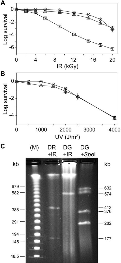
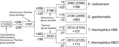
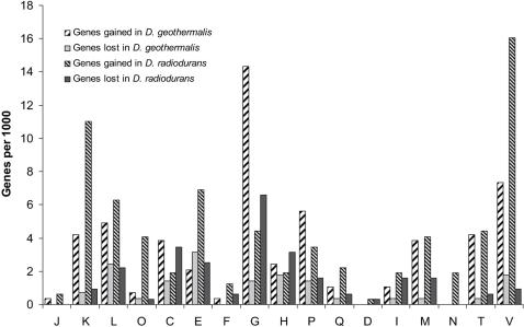
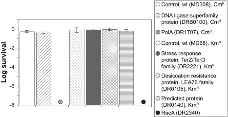
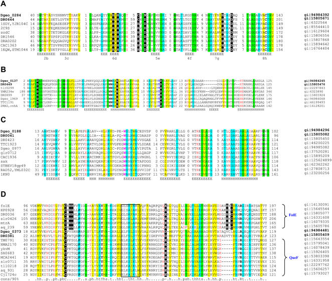
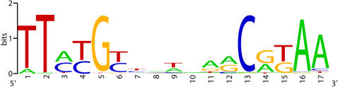
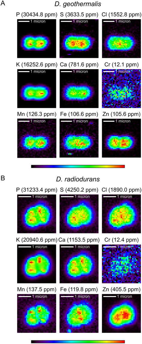
Bacteria of the genus Deinococcus are extremely resistant to ionizing radiation (IR), ultraviolet light (UV) and desiccation. The mesophile Deinococcus radiodurans was the first member of this group whose genome was completely sequenced. Analysis of the genome sequence of D. radiodurans, however, failed to identify unique DNA repair systems. To further delineate the genes underlying the resistance phenotypes, we report the whole-genome sequence of a second Deinococcus species, the thermophile Deinococcus geothermalis, which at its optimal growth temperature is as resistant to IR, UV and desiccation as D. radiodurans, and a comparative analysis of the two Deinococcus genomes. Many D. radiodurans genes previously implicated in resistance, but for which no sensitive phenotype was observed upon disruption, are absent in D. geothermalis. In contrast, most D. radiodurans genes whose mutants displayed a radiation-sensitive phenotype in D. radiodurans are conserved in D. geothermalis. Supporting the existence of a Deinococcus radiation response regulon, a common palindromic DNA motif was identified in a conserved set of genes associated with resistance, and a dedicated transcriptional regulator was predicted. We present the case that these two species evolved essentially the same diverse set of gene families, and that the extreme stress-resistance phenotypes of the Deinococcus lineage emerged progressively by amassing cell-cleaning systems from different sources, but not by acquisition of novel DNA repair systems. Our reconstruction of the genomic evolution of the Deinococcus-Thermus phylum indicates that the corresponding set of enzymes proliferated mainly in the common ancestor of Deinococcus. Results of the comparative analysis weaken the arguments for a role of higher-order chromosome alignment structures in resistance; more clearly define and substantially revise downward the number of uncharacterized genes that might participate in DNA repair and contribute to resistance; and strengthen the case for a role in survival of systems involved in manganese and iron homeostasis.


Conflict of interest statement

Competing Interests: The authors have declared that no competing interests exist.

Figures

Figure 1
Figure 1. Radiation resistance and genome structure of D. geothermalis and D. radiodurans. A, IR (60Co, 5.5 kGy/h).
B, UV (254 nm) (3 J/m2 s−1). Open circle, D. radiodurans (32°C); open triangle, D. geothermalis (50°C); and open square, D. geothermalis (32°C). Values are from three independent trials with standard deviations shown. At near-optimal growth temperatures, the 10% survival values (D10) following IR for D. radiodurans (32°C) and D. geothermalis (50°C) are 15 kGy; for E. coli, 0.7 kGy (37°C) ; and for T. thermophilus (HB27) 0.8 kGy (65°C) . C, PFGE of genomic DNA prepared from irradiated (0.2 kGy) D. radiodurans (DR+IR) and D. geothermalis (DG+IR); and genomic DNA from non-irradiated D. geothermalis digested with SpeI (DG+SpeI). (M) PFGE DNA size markers. PFGE was as described previously .
Figure 2
Figure 2. Whole genome evolutionary reconstructions for D. radiodurans, D. geothermalis, T. thermophilus (HB8) and T. thermophilus (HB27).
For each internal node of tree (open boxes), the inferred number of tdCOGs is shown. For each tree branch the inferred number of tdCOGs lost (minus sign) and gained (plus sign) is shown. For the deep ancestor of the Cyanobacteria, Actinobacteria and Deinococcus-Thermus group (shaded box), the inferred number of COGs is shown. For the extant species, the number of tdCOGs, the number of proteins in tdCOGs (in parentheses), and the number of “free” (not assigned to tdCOGs) proteins (plus sign) are shown.
Figure 3
Figure 3. Gene-gain and gene-loss for different functional groups for D. radiodurans and D. geothermalis.
Designations of functional groups (from the COG database): J–Translation, ribosomal structure and biogenesis; K–Transcription; L–DNA replication, recombination and repair; D–Cell division and chromosome partitioning; O–Posttranslational modification, protein turnover, chaperones; M–Cell envelope and outer membrane biogenesis; N–Cell motility and secretion; P–Inorganic ion transport and metabolism; T–Signal transduction mechanisms; C–Energy production and conversion; G–Carbohydrate transport and metabolism; E–Amino acid transport and metabolism; F–Nucleotide transport and metabolism; H–Coenzyme metabolism; I–Lipid metabolism; Q–Secondary metabolites biosynthesis, transport and catabolism; V–genes involved in stress response and microbial defense.
Figure 4
Figure 4. IR resistance of wild-type (ATCC BAA-816) and D. radiodurans mutants lacking orthologs in D. geothermalis (DSM 11300).
Survival values following 9 kGy (60Co) are from three independent trials with standard deviations shown. The structure of the homozygous mutants DRB0100, DR2221, DR105 and DR0140 are presented in Figure S6.
Figure 5
Figure 5. Multiple alignments of selected families conserved in two Deinococcus species.
The multiple alignments were constructed for selected representative sets of sequences by the MUSCLE program . Where necessary, alignments were modified manually on the basis of PSI-BLAST outputs . The positions of the first and the last residue of the aligned region in the corresponding protein are indicated for each sequence. The numbers within the alignment refer to the length of inserts that are poorly conserved between all the families. Secondary structure elements are denoted as follows: E-β-strand; and H-α-helix. The coloring scheme is as follows: predominantly hydrophobic residues are high-lighted in yellow; positions with small residues are in green; positions with turn-promoting residues are in cyan; positions with polar residues are in red; hydroxyl-group containing residues are in blue; aromatic residues are in magenta; and invariant, highly conserved groups are in boldface. A, DR0644-Dgeo_0284 conserved pair of orthologs belong to the copper/Zinc superoxide dismutase family; shaded letters refer to amino acids that play an important role in the Cu2+/Zn2+ coordination environment and in the active site region. The bottom line shows the correspondence between the most conserved regions corresponding to the β-stand structural core and conserved in most family members as denoted in Bordo et al . B, Dgeo_0137-DR0449 are highly specific for the Deinococcus lineage proteins that have an RNAse H-related domain. Catalytic residues conserved in the RNAse H family are shaded. Secondary structure elements are shown for E. coli RNase HI (PDB:2rn2). C, DR0041-Dgeo_0188 is another conserved pair (DdrA-related) of proteins belonging to the Rad52 family of DNA single-strand annealing proteins . Secondary structure elements are shown for human RAD52 (PDB:1KN0) . sak is a phage gene described previously ; D, DR0381-Dgeo_0373 are diverged homologs of NADPH-dependent nitrile reductase (GTP cyclohydrolase I family) that might be involved in nucleotide metabolism. The conserved Cys and Glu found in the substrate binding pocket of both protein families and specific zinc-binding and catalytic residues in the FolE family are shaded. The QueF family motif is boxed. Other catalytic residues in FolE not found in QueF are in yellow. Genbank Identifier (gi) numbers are listed on the right.
Figure 6
Figure 6. Sequence signature of a predicted site of a radiation response regulator.
Four different nucleotides are shown by four letters (A, G, C, T) in different colors. The height of the letter is proportional to its contribution to the information content in the corresponding position of the multiple alignment used for “sequence logo” construction. The figure was constructed by the “sequence logo” program described previously .
Figure 7
Figure 7. X-ray fluorescence (XRF) microprobe element distribution maps .
A, D. geothermalis (diplococcus). B, D. radiodurans (tetracocus). Cells were harvested from mid-logarithmic cultures in undefined rich medium, imaged, and quantified as described previously . The element distribution images are plotted to different scales designated by a single color-box, where red represents the highest concentration and black the lowest. ppm values in parentheses next to the element symbol correspond to red. XRF microprobe analysis measurements were made at beamline 2ID-D at the Advanced Photon Source, Argonne National Laboratory as described recently .

Similar articles

Cited by

References

    1. Gupta RS. Protein phylogenies and signature sequences: A reappraisal of evolutionary relationships among archaebacteria, eubacteria, and eukaryotes. Microbiol Mol Biol Rev. 1998;62:1435–1491. - PMC - PubMed
    1. Wolf YI, Rogozin IB, Grishin NV, Tatusov RL, Koonin EV. Genome trees constructed using five different approaches suggest new major bacterial clades. BMC Evol Biol. 2001;1:8. - PMC - PubMed
    1. Lai WA, Kampfer P, Arun AB, Shen FT, Huber B, et al. Deinococcus ficus sp. nov., isolated from the rhizosphere of Ficus religiosa L. Int J Syst Evol Microbiol. 2006;56:787–791. - PubMed
    1. Gutman PD, Fuchs P, Minton KW. Restoration of the DNA damage resistance of Deinococcus radiodurans DNA polymerase mutants by Escherichia coli DNA polymerase I and Klenow fragment. Mutat Res. 1994;314:87–97. - PubMed
    1. Daly MJ, Gaidamakova EK, Matrosova VY, Vasilenko A, Zhai M, et al. Accumulation of Mn(II) in Deinococcus radiodurans facilitates gamma-radiation resistance. Science. 2004;306:1025–1028. - PubMed

Publication types