. 2007 Sep 26;2(9):e956.
doi: 10.1371/journal.pone.0000956.

MVB-12, a fourth subunit of metazoan ESCRT-I, functions in receptor downregulation

Affiliations

MVB-12, a fourth subunit of metazoan ESCRT-I, functions in receptor downregulation

Anjon Audhya et al. PLoS One. .

Abstract

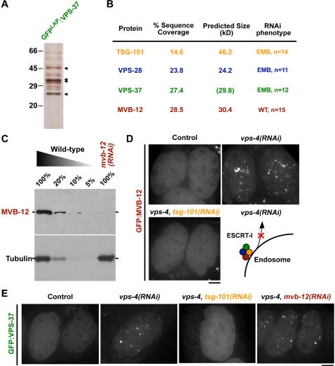
After ligand binding and endocytosis, cell surface receptors can continue to signal from endosomal compartments until sequestered from the cytoplasm. An important mechanism for receptor downregulation in vivo is via the inward budding of receptors into intralumenal vesicles to form specialized endosomes called multivesicular bodies (MVBs) that subsequently fuse with lysosomes, degrading their cargo. This process requires four heterooligomeric protein complexes collectively termed the ESCRT machinery. In yeast, ESCRT-I is a heterotetrameric complex comprised of three conserved subunits and a fourth subunit for which identifiable metazoan homologs were lacking. Using C. elegans, we identify MVB-12, a fourth metazoan ESCRT-I subunit. Depletion of MVB-12 slows the kinetics of receptor downregulation in vivo, but to a lesser extent than inhibition of other ESCRT-I subunits. Consistent with these findings, targeting of MVB-12 to membranes requires the other ESCRT-I subunits, but MVB-12 is not required to target the remaining ESCRT-I components. Both endogenous and recombinant ESCRT-I are stable complexes with a 1:1:1:1 subunit stoichiometry. MVB-12 has two human homologs that co-localize and co-immunoprecipitate with the ESCRT-I component TSG101. Thus, MVB-12 is a conserved core component of metazoan ESCRT-I that regulates its activity during MVB biogenesis.


Conflict of interest statement

Competing Interests: The authors have declared that no competing interests exist.

Figures

Figure 1
Figure 1. ESCRT-I components mediate degradation of the cell surface protein GFP:CAV-1 after its internalization.
(A) Table listing the known subunits of ESCRT-I in S. cerevisiae, H. sapiens and C. elegans. (B) Schematic of the reproductive system of the adult C. elegans hermaphrodite. Oocytes (blue) aligned in a row are fertilized in a sequential conveyor belt-like fashion with one ovulation event occurring ∼every 20 minutes . Pre-fertilization oocytes are numbered -1, -2, etc. with respect to their position relative to the spermatheca. Similarly, fertilized embryos (green), which are at progressively later stages in development as their distance from the spermatheca increases, are numbered +1, +2, +3, etc. (C) Spinning disk confocal optics were used to image anesthetized control (n = 14), tsg-101(RNAi) (n = 15), vps-28(RNAi) (n = 11), and vps-37(RNAi) (n = 10) adult hermaphrodites expressing GFP:CAV-1. Both differential interference contrast (DIC, left) and fluorescence (right) images are shown. Schematics in the center column are traces of the DIC images in which the location of the spermatheca (red), the unfertilized oocytes (blue) and the embryos (green) are highlighted. Boxes indicate the presence of embryos at or beyond the two-cell stage, where GFP:CAV-1 is normally absent. Scale bar is 25 µm.
Figure 2
Figure 2. Identification of a fourth C. elegans ESCRT-I subunit. (A) A fusion of VPS-37 with a GFP containing tandem affinity purification tag (GFPLAP:VPS-37) was isolated from embryo extracts by immunoprecipitation with anti-GFP antibodies.
After protease cleavage to remove the GFP tag, proteins were re-isolated on S-protein agarose and eluted. A silver stained gel of the eluted proteins is shown. Arrowheads indicate the predicted mobility of the proteins identified by mass spectrometry. (B) Table summarizing the percent sequence coverage, predicted molecular weight, and RNAi phenotype for each of the four specific proteins in the eluate identified by solution mass spectrometry. For VPS-37, the predicted size including the additional mass of the affinity purification tag is shown in parentheses. In addition to the four proteins shown, a common contaminant (LEV-11) was also identified by mass spectrometry, but is not included in the table. (C) Western blots of extracts prepared from wild type worms or worms specifically depleted of MVB-12 by RNAi. Serial dilutions of extract prepared from untreated worms were loaded to quantify depletion levels. (D) Spinning disk confocal optics were used to image GFP:MVB-12 in control (n = 15), vps-4(RNAi) (n = 13), and vps-4, tsg-101(RNAi) (n = 15) embryos in utero. Schematic illustrates the effect of depleting VPS-4, the AAA-ATPase required to recycle endosome-associated ESCRT-I, returning it to the cytoplasm (lower right). Scale bar is 10 µm. (E) Spinning disk confocal optics were used to image GFP:VPS-37 in control (n = 10), vps-4(RNAi) (n = 8), vps-4, tsg-101(RNAi) (n = 9), and vps-4, mvb-12(RNAi) (n = 9) embryos in utero. Scale bar is 10 µm.
Figure 3
Figure 3. ESCRT-I is a 1∶1∶1∶1 heterotetrameric complex both in vivo and in vitro. The results presented in each panel are representative of three individual experiments performed. Schematics in the upper right corner of each panel indicate the ESCRT-I components present in each experiment.
(A) TSG-101:6xHIS, VPS-28, and VPS-37 were co-expressed and purified from E. coli extracts using nickel resin. A Coomassie stained gel of the peak elution fractions after fractionation of the recombinant complex on a Superose 6 gel filtration column is shown. The intensities of each band were measured to identify the peak, and a Stokes radius was calculated for each protein based on comparison with the elution profiles of known standards. The asterisk highlights a breakdown product of TSG-101. (B) The recombinant complex described in A, was fractionated on a 5–20% sucrose gradient. A western blot of the fractions for TSG-101 is shown. The asterisk highlights a breakdown product of TSG-101. The intensities of each band were measured to identify the peak, and S-values were calculated for each protein based on the location of known standards run on a parallel gradient. (C) TSG-101:6×HIS, VPS-28, VPS-37 and MVB-12 were co-expressed and purified from E. coli extracts using nickel resin. A Coomassie stained gel of the peak elution fractions after fractionation of the recombinant complex on a Superose 6 gel filtration column is shown. The Stokes radius of the complex, calculated as described for A, is indicated. (D) The recombinant complex described in C, was fractionated on a 5–20% sucrose gradient. A western blot of the fractions for MVB-12 is shown. The S-value of the complex, calculated as described in B is indicated. (E) Western blots of embryo extracts fractionated on a Superose 6 gel filtration column. The peaks corresponding to TSG-101 and MVB-12 are largely overlapping. The Stokes radius of the complex, calculated as described for A, is indicated. (F) Western blots of embryo extracts after fractionation on a 5–20% sucrose density gradient. TSG-101 and MVB-12 co-fractionate, both migrating with an S-value close to 5.0. The S-value of the complex, calculated as described in B is shown.
Figure 4
Figure 4. Depletion of MVB-12 slows the degradation of internalized RME-2, but to a lesser extent than inhibition of the ESCRT-I component TSG-101.
(A) Spinning disk confocal optics were used to image control (n = 7), mvb-12(RNAi) (n = 8), and tsg-101(RNAi) (n = 6) embryos co-expressing RME-2:GFP and RFP:RAB-5 in utero. Representative sections are shown. Times are in minutes relative to oocyte ovulation. Arrowheads highlight the transient accumulation of RME-2:GFP on the cell surface at the 10 min timepoint. Scale bar is 10 µm. (B) Higher magnification (2x) views of a portion of the control images acquired in (A). At early timepoints after RME-2:GFP internalization, the GFP signal co-localizes with RFP:RAB-5. At later timepoints, both the extent to which the localization of RME-2:GFP is co-incident with that of RFP:RAB-5 and the overall intensity of the RME-2:GFP signal are reduced. Scale bar is 5 µm. (C) The distribution of the time (in minutes after ovulation) when the RME-2:GFP signal was no longer detectable at the cell surface is shown for control, mvb-12(RNAi), and tsg-101(RNAi) embryos. (D) The distribution of the time (in minutes after ovulation) when >90% of RME-2:GFP containing endosomes were no longer positive for RFP:RAB-5 is shown for control, mvb-12(RNAi), and tsg-101(RNAi) embryos. GFP-positive and RFP-positive endosomes with signals above the cytoplasmic background were identified using individual thresholds. At 1 min intervals, each GFP-labeled endosome was examined for RFP signal. The center of the first 5 min time interval over which less than 10% of the total GFP-positive endosomes also had a detectable RFP signal is plotted for each embryo. (E) The distribution of the time (in minutes after ovulation) when >90% of the RME-2:GFP fluorescence present 10 minutes post ovulation had been lost is shown for control, mvb-12(RNAi), and tsg-101(RNAi) embryos. The total integrated RME-2:GFP fluorescence intensity was measured in a 10 µm×10 µm box for each timepoint and the time when this value fell below 10% of the total fluorescence in an identical box at the 10 min timepoint in the same embryo is plotted. Background integrated fluorescence intensity measured in an identical box in older (>50 cell stage) control embryos was subtracted before calculating the percentage of fluorescence remaining.
Figure 5
Figure 5. The human MVB-12 homologs, MVB12A and MVB12B, are new components of human ESCRT-I.
(A) Sequence alignment of the C. elegans protein with MVB12A and MVB12B was performed using ClustalW. Amino acids that are identical between two of the three sequences are highlighted in light blue and between all three sequences in dark blue. (B) HeLa cells transiently transfected with GFP:MVB12B were detergent extracted, fixed and stained with antibodies to GFP and human TSG101 (n = 23). A single section from a representative 3D computationally deconvolved data set is shown. A color overlay of GFP:MVB12B (red) and TSG101 (green) and a higher magnification (8x) view of the indicated boxed region are also shown. Scale bar is 10 µm. (C) HeLa cells transiently transfected with RFP:MVB12B and either wild type hVPS4B:GFP (n = 13) or mutant hVPS4BE235Q:GFP (n = 21) were imaged using spinning disk confocal microscopy. Representative color overlays of RFP:MVB12B (red) and hVPS4:GFP (green) are also shown. Scale bar is 10 µm. (D) Immunoprecipitations with antibodies to GFP, or GST as a control, were performed on extracts prepared from HeLa cells transfected with either GFP:MVB12A or GFP:MVB12B. Western blots of the starting extracts (load, 10% of total) and the control (10% of total) and GFP (10% of total) immunoprecipitates were probed with antibodies to TSG101.

References

    1. Schlessinger J. Cell signaling by receptor tyrosine kinases. Cell. 2000;103:211–225. - PubMed
    1. Vivekanand P, Rebay I. Intersection of signal transduction pathways and development. Annu Rev Genet. 2006;40:139–157. - PubMed
    1. Bache KG, Slagsvold T, Stenmark H. Defective downregulation of receptor tyrosine kinases in cancer. EMBO J. 2004;23:2707–2712. - PMC - PubMed
    1. Porter AC, Vaillancourt RR. Tyrosine kinase receptor-activated signal transduction pathways which lead to oncogenesis. Oncogene. 1998;17:1343–1352. - PubMed
    1. Katzmann DJ, Odorizzi G, Emr SD. Receptor downregulation and multivesicular-body sorting. Nat Rev Mol Cell Biol. 2002;3:893–905. - PubMed

Publication types

LinkOut - more resources