. 2007 Sep 26;2(9):e959.
doi: 10.1371/journal.pone.0000959.

The INT6 cancer gene and MEK signaling pathways converge during zebrafish development

Affiliations

The INT6 cancer gene and MEK signaling pathways converge during zebrafish development

Michal Grzmil et al. PLoS One. .

Abstract

Background: Int-6 (integration site 6) was identified as an oncogene in a screen of tumorigenic mouse mammary tumor virus (MMTV) insertions. INT6 expression is altered in human cancers, but the precise role of disrupted INT6 in tumorigenesis remains unclear, and an animal model to study Int-6 physiological function has been lacking.

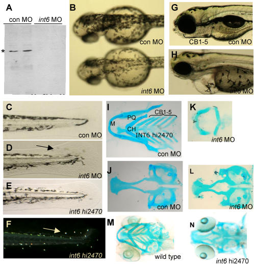
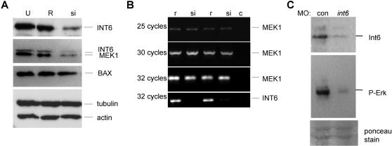
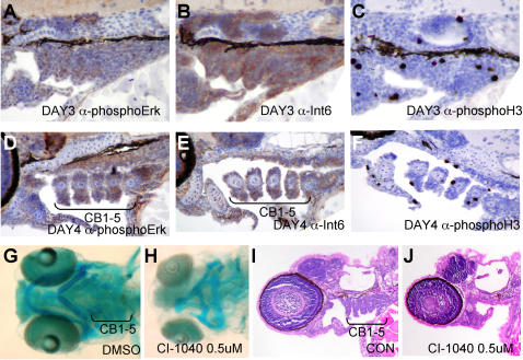
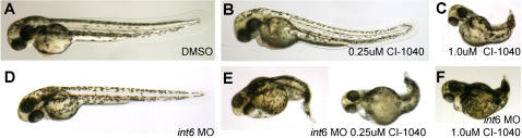
Principal findings: Here, we create an in vivo model of Int6 function in zebrafish, and through genetic and chemical-genetic approaches implicate Int6 as a tissue-specific modulator of MEK-ERK signaling. We find that Int6 is required for normal expression of MEK1 protein in human cells, and for Erk signaling in zebrafish embryos. Loss of either Int6 or Mek signaling causes defects in craniofacial development, and Int6 and Erk-signaling have overlapping domains of tissue expression.

Significance: Our results provide new insight into the physiological role of vertebrate Int6, and have implications for the treatment of human tumors displaying altered INT6 expression.


Conflict of interest statement

Competing Interests: The authors have declared that no competing interests exist.

Figures

Figure 1
Figure 1. Int6 is essential for zebrafish embryonic development.
(A) Western blot analysis of Int6 (*) in zebrafish embryos injected with a control (con) MO or int6 MO. (B) int6 morphant melanocytes are less darkly pigmented. (C–F) Int6 is required for pigment cell placement in the tail, as int6 morphants and int6hi2470 mutants have misplaced pigment cells in the tail fin (D, arrow). Ambient light illuminates the iridophore ‘star-light’ pattern seen in the int6hi2470 embryos (F, arrow). (G–H) By 5 dpf, embryos injected with a con MO have clearly visible certobranical arches, while int6 morphants do not have visible certobranical arches, in addition to other abnormalities, including unconsumed yolk sac, heart and eye development. (I–N). Alcian blue staining of 5 dpf embryos shows loss of ceratobrancial arches 1 through 5. M, Meckel's; PQ, palatoquadrate; CH, ceratohyal; CB, ceratobrancial.
Figure 2
Figure 2. INT6 is required for MEK protein levels and Erk-signaling.
A. Osteosarcoma U2-OS cells untransfected (U), or transfected with siRNA targeted against the INT6 mRNA (si) or the reverse sequence (R), show reduced levels of INT6 and MEK1 protein specifically after transfection with INT6-siRNA, but no reduction in BAX, tubulin or actin protein levels. (B) Semi-quantitative-PCR shows MEK1 mRNA is unaffected in reverse sequence and INT6-siRNA treated cells, coupled with the expected reduced levels of the INT6 message in the INT6-siRNA transfected cells. c, PCR control without DNA. (C) Phospho-Erk levels are reduced in int6 morphants, while ponceau stain detects equal loading of protein on the gel.
Figure 3
Figure 3. Int6 and phospho-Erk expression in the developing zebrafish embryo, and pharmacological inhibition of Mek alters ceratobrancial (CB) arches.
(A–F). Immuno-histochemistry of Int6 and phospho-Erk in the developing craniofacial tissues, counter stained with hematoxylin, and phospho-histone H3 to show cycling cells. (G, H) Ventral whole mount views of Alcian blue stained pharyngeal cartilages show loss of ceratobrancial arches 1–5 and a reduction of Meckel's (M), palatoquadrate (PQ) and ceratohyal (CH) cartilages in 4 dpf embryos treated with 0.5 uM CI-1040. (I, J) Sections of 4 dpf embryos hematoxylin and eosin stained after 0.5 uM CI-1040 treatment reveals loss of CB arches 1–5 (brackets). E, Ethmoid plate; PC, Parachordal cartilage.
Figure 4
Figure 4. Int6 and Mek signaling interact in vivo.
(A–C). Only embryos treated with the 1.0 µM CI-1040, and not 0.25 µM, show a loss of posterior structures, in contrast to (D) int6 morphants (int6 MO 0.25 ng). (E, F) In combination with 0.25 µM of CI-1040, 0.25 µg of int6 MO causes a dramatic alteration of the anterior-posterior axis.

References

    1. Callahan R, Smith GH. MMTV-induced mammary tumorigenesis: gene discovery, progression to malignancy and cellular pathways. Ongogene. 2000;19:992–1001. - PubMed
    1. Nusse R, Varmus HE. Many Tumors Induced by the Mouse Mammary Tumor Virus Contain a Provirus Integrated in the Same Region of the Host Genome. Cell. 1982;31:99–109. - PubMed
    1. Rijsewijk F, Schuermann M, Wagenaar E, Parren P, Weigel D, et al. The Drosophila homolog of the mouse mammary oncogene int-1 is identical to the segment polarity gene wingless. Cell. 1987;50:649–657. - PubMed
    1. Clevers H. Wnt/ß-Catenin Signaling in Development and Disease. Cell. 2006;3:469–480. - PubMed
    1. Tekmal RR, Keshava N. Role of MMTV integration locus cellular genes in breast cancer. Front Biosci. 1997;379:519–526. - PubMed

Publication types

MeSH terms