Clinical and diagnostic imaging findings predict anesthetic complications in children presenting with malignant mediastinal masses
- PMID: 17897276
- PMCID: PMC4400730
- DOI: 10.1111/j.1460-9592.2007.02279.x
Clinical and diagnostic imaging findings predict anesthetic complications in children presenting with malignant mediastinal masses
Abstract
Background: The presence of a mediastinal mass in a child poses significant anesthesia-related risks including death. To optimize outcome clinicians must be able to predict which patients are at highest risk of anesthetic complications.
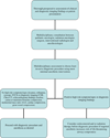
Methods: We conducted a retrospective review of 118 pediatric patients who presented with mediastinal masses. We investigated their medical records for clinical symptoms and signs at presentation and reviewed their chest radiographs, computed tomography scans, and echocardiograms and electrocardiograms when available. We then conducted analyses to identify clinical and diagnostic imaging features associated with anesthesia-related complications.
Results: Eleven of 117 [9.4%, 95% confidence interval (CI) 4.1-14.7%] patients experienced an anesthesia-related complication. Four preoperative features were significantly associated with anesthetic complications: orthopnea (P = 0.033, odds ratio (OR) 5.31, 95% CI, 1.15-24.56), upper body edema (P = 0.035, OR 8.00, 95% CI, 1.16-55.07), great vessel compression (P = 0.037, OR 5.41, 95% CI, 1.11-26.49), and main-stem bronchus compression (P = 0.044, OR 5.11, 95% CI, 1.05-24.92). The presence of pleural effusion (P = 0.060, OR 4.53, 95% CI, 0.94-21.96) or tracheal compression (P = 0.061, OR 5.09, 95% CI, 0.93-27.81) also appeared to be risk factors. Although the rate of anesthesia-related complications detected in our cohort was comparable with that found in earlier studies, the events were less severe.
Conclusions: Patients who present with orthopnea, upper body edema, great vessel compression and main stem bronchus compression are at risk of anesthesia-related complications. The low severity of complications in our series may reflect a combination of factors: use of the least invasive method such as interventional radiology to obtain tissue for diagnosis, completion of a thorough preoperative assessment and minimal anesthesia intervention.
Figures
References
-
- Levin H, Bursztein S, Heifetz M. Cardiac arrest in a child with an anterior mediastinal mass. Anesth Analg. 1985;64:1129–1130. - PubMed
-
- Northrip DR, Bohman BK, Tsueda K. Total airway occlusion and superior vena cava syndrome in a child with an anterior mediastinal tumor. Anesth Analg. 1986;65:1079–1082. - PubMed
-
- Keon TP. Death on induction of anesthesia for cervical node biopsy. Anesthesiology. 1981;55:471–472. - PubMed
-
- Bray RJ, Fernandes FJ. Mediastinal tumour causing airway obstruction in anaesthetised children. Anaesthesia. 1982;37:571–575. - PubMed
-
- Prakash UB, Abel MD, Hubmayr RD. Mediastinal mass and tracheal obstruction during general anesthesia. Mayo Clin Proc. 1988;63:1004–1011. - PubMed
Publication types
MeSH terms
Grants and funding
LinkOut - more resources
Full Text Sources
Other Literature Sources
Medical


