. 2007 Sep 26:7:61.
doi: 10.1186/1472-6750-7-61.

pHUSH: a single vector system for conditional gene expression

Affiliations

pHUSH: a single vector system for conditional gene expression

Daniel C Gray et al. BMC Biotechnol. .

Abstract

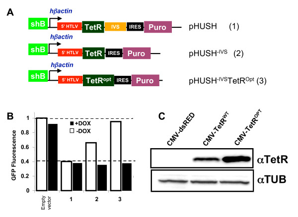
Background: Conditional expression vectors have become a valuable research tool to avoid artefacts that may result from traditional gene expression studies. However, most systems require multiple plasmids that must be independently engineered into the target system, resulting in experimental delay and an increased potential for selection of a cell subpopulation that differs significantly from the parental line. We have therefore developed pHUSH, an inducible expression system that allows regulated expression of shRNA, miRNA or cDNA cassettes on a single viral vector.

Results: Both Pol II and Pol III promoters have been successfully combined with a second expression cassette containing a codon-optimized tetracycline repressor and selectable marker. We provide examples of how pHUSH has been successfully employed to study the function of target genes in a number of cell types within in vitro and in vivo assays, including conditional gene knockdown in a murine model of brain cancer.

Conclusion: We have successfully developed and employed a single vector system that enables Doxycycline regulated RNAi or transgene expression in a variety of in vitro and in vivo model systems. These studies demonstrate the broad application potential of pHUSH for conditional genetic engineering in mammalian cells.


Figures

Figure 1
Figure 1
Luciferase-based transient co-transfection experiments to compare shRNA and miRNA design schemes. Knockdown studies were performed in duplicate as co-transfections of shRNA:pGL3-gene:pRL at the indicated ratio. Firefly luciferase values are divided by renilla values to control for transfection efficiency, and all knockdown data are normalized to control hairpins. (A) Comparison of published modifications to standard H1-shRNA design (F = frayed, miR23 = loop derived from miR-23, and N19 = standard pSuper shRNA design) directed against the pGL3-MELK reporter. Cells were transfected with a 10:1 ratio of the indicated shRNA to pGL3-Melk reporter. (B) CMV-miR to H1-shRNA conversion scheme. The active 'siRNA' was identified within the pol II-miRNA and converted to a standard H1-shRNA hairpin (black bars = Drosha and Dicer processing; red = guide strand, blue = miR-155 loop, pink = pSuper loop). (C) Comparison of H1-shRNA and CMV-miR vectors corresponding to murine p53 sequences transfected at 7.5:1, 5:1 and 2.5:1 (shRNA:pGL3-mu-p53). (D) The removal of the EmGFP leader renders miR-p53-1 non-functional at a co-transfection ratio of 10:1.
Figure 2
Figure 2
pHUSH vector design and optimization (A) Vector diagrams of the pHUSH vector series: (1) original pHUSH backbone (2) pHUSH-IVS in which the synthetic intron sequence between the TetR ORF and the IRES is removed, and (3) pHUSH-IVSTetROpt with a codon-optimzed TetR (B) Increased TetR expression ensures regulation of GFP-Melk fusion protein in the absence of doxycycline. HEK 293 cells expressing a GFP-MELK fusion were transfected with the pHUSH vector series described above all containing the shB Melk targeting shRNA. After selection with 3 μg/ml puromycin, the resulting stable pools were cultured in the absence (open bars) or presence (filled bars) of 1 μg/ml doxycycline for five days and GFP-Melk expression analyzed by FACS. Data is normalized to the mean fluorescence intensity (10,000 acquired events) for cells containing a pHUSH empty vector control. A representative experiment is shown. (C) Codon optimization of TetR open reading frame (ORF) increases translation. Both the original (WT) and codon-optimized (OPT) TetR ORFs were transiently expressed in 293T cells. Forty-eight hours post-transfection, cell lysates were prepared and Western blotted with an anti-TetR antibody and an anti-tubulin antibody.
Figure 3
Figure 3
Multiple TetO2 operons within the Pol III promoter enhances regulated knock-down. (A) 293T cells were transfected with 15 ng of pGL3-huMelk in the presence of decreasing molar ratios of TetR and the appropriate H1-shRNA vectors using the indicated molar ratio of TetR:H1-shRNA vector. The maximal level of gene knockdown that can be observed with either promoter is represented by cells transfected with the appropriate H1-shRNA vector and no TetR (0:1). Both H1-shRNA constructs expressed the shB Melk targeting shRNA. Luciferase expression was measured as described in the Methods 48 hours post transfection. (B) Dox regulated Melk knockdown is maintained in differentiated ES cells with the 2 × TetO2 promoter configuration. Embryoid bodies were generated as described in the Methods from stable pHUSH ES cell clones with shRNA targeting MELK or Luciferase. shMELK ES cell lines were generated with either one TetO2 (1×-TetO2, colony A6) or two TetO2 (2×-TetO2, colony 3C11) operons (see also additional file 3). ES cells lines were differentiated into embryoid bodies in the presence or absence of 1 ug/ml doxycycline and the level of Melk expression determined by qRT-PCR and normalized to the housekeeping gene SPF31 (RefSeq NM_014280).
Figure 4
Figure 4
Retroviral delivery of the pHUSH system allows the inducible expression of shRNA, miRNA or proteins. (A) Vector diagram of the pHUSH retroviral system. Inducible pol II or pol III expression cassettes are introduced by Gateway® recombination. (B) Improved p53 knockdown by CMV-miRNA in comparison to H1-shRNA vectors at low MOI. H1-shRNA and CMV-miRNA vectors with hairpins designed against murine p53 as described in additional file 2 were cloned into the pHUSH retroviral backbone. 3T3 cells were infected at an MOI = 0.2, selected in 1.5 ug/mL puromycin, and cultured ± 1 ug/mL Dox, Knockdown of p53 was determined by qRT-PCR at Day 4 post-Dox and was normalized to p53 levels in the absence of Dox. (C) Inducible expression of proteins by the pHUSH retroviral system. Inducible EGFP and EGFP-MELK-D150A expression cassettes were cloned into the pHUSH retroviral backbone. HCT116 cells were infected with the appropriate viral construct, selected in 5 ug/mL puromycin, and treated ± 1 ug/mL Dox for 72 h. Equal total protein was fractionated by non-reducing SDS-PAGE and imaged by the Typhoon fluorescent imager at the indicated excitation and emission spectra (WB = western blot). Equal loading was confirmed by western blotting with an anti-tubulin antibody.
Figure 5
Figure 5
Effective in vivo silencing of luciferase in subcutaneous and intracranial tumor models. (A) Mean intensity luciferase expression indicates Dox induced luciferase knockdown in subcutaneous tumor xenograft at day 3 and 5 post-injection. Eight animals are represented for each treatment condition. (B) Dox treatment or luciferase knockdown does not reduce tumor growth of SVT2 cells in SCID mice. (C) Mean intensity luciferase expression demonstrates Dox-induced luciferase knockdown relative to controls at days 3 (9.5 fold), 5 (13.8 fold) and 9 (11 fold) post-injection. The luminescence values for each time point have been normalized to the respective intensity value at day 0. Five animals are represented for each treatment condition. Representative BLI images are shown in additional file 7.
Figure 6
Figure 6
Robust and titratable depletion of oncogenic B-Raf in melanoma tumor lines elicits the expected phenotypes in vitro and in vivo. (A) Knockdown of BRAF at Day 2 upon Dox addition as determined by western blotting in A375 cells. BRAF knockdown results in a significant reduction in phospho-ERK levels. (B) Proliferation assay reveals a growth stasis phenotype upon BRAF knockown in comparison to a shEGFP control in A375 cells. (C) Dose dependent decrease in VEGF secretion correlates with BRAF knockdown 24 and 48 h post Dox addition. (D) In vivo BRAF knockdown detected by non-invasive bioluminescence imaging. A previously characterized LOX-IMV1 clone containing pHUSH-Braf-shRNA [26] was re-engineered to express a Luc-BRAF transcipt fusion. After initiating a subcutaneous tumor model in Scid-beige mice, the expression level of a Braf targeting shRNA was indirectly monitored by bioluminescence imaging at 0, 0.01 mg/mL, 0.02 mg/mL, and 0.5 mg/mL Dox. (E) Day 25 tumors were harvested and equal total protein was assayed for luciferase enzyme activity in vitro.

References

    1. Elbashir SM, Harborth J, Lendeckel W, Yalcin A, Weber K, Tuschl T. Duplexes of 21-nucleotide RNAs mediate RNA interference in cultured mammalian cells. Nature. 2001;411:494–498. doi: 10.1038/35078107. - DOI - PubMed
    1. Brummelkamp TR, Bernards R, Agami R. A system for stable expression of short interfering RNAs in mammalian cells. Science. 2002;296:550–553. doi: 10.1126/science.1068999. - DOI - PubMed
    1. Paddison PJ, Caudy AA, Bernstein E, Hannon GJ, Conklin DS. Short hairpin RNAs (shRNAs) induce sequence-specific silencing in mammalian cells. Genes Dev. 2002;16:948–958. doi: 10.1101/gad.981002. - DOI - PMC - PubMed
    1. Sandy P, Ventura A, Jacks T. Mammalian RNAi: a practical guide. Biotechniques. 2005;39:215–224. - PubMed
    1. Silva J, Chang K, Hannon GJ, Rivas FV. RNA-interference-based functional genomics in mammalian cells: reverse genetics coming of age. Oncogene. 2004;23:8401–8409. doi: 10.1038/sj.onc.1208176. - DOI - PubMed

MeSH terms

Substances

LinkOut - more resources