Comparative Study
. 2007 Sep 26:8:79.
doi: 10.1186/1471-2202-8-79.

Dynamic remodelling of synapses can occur in the absence of the parent cell body

Affiliations
Comparative Study

Dynamic remodelling of synapses can occur in the absence of the parent cell body

Natalia L Bettini et al. BMC Neurosci. .

Abstract

Background: Retraction of nerve terminals is a characteristic feature of development, injury and insult and may herald many neurodegenerative diseases. Although morphological events have been well characterized, we know relatively little about the nature of the underlying cellular machinery. Evidence suggests a strong local component in determining which neuronal branches and synapses are lost, but a greater understanding of this basic neurological process is required. Here we test the hypothesis that nerve terminals are semi-autonomous and able to rapidly respond to local stimuli in the absence of communication with their parent cell body.

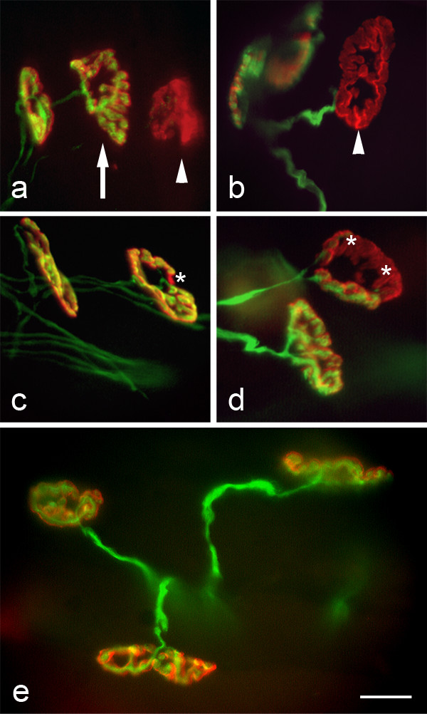
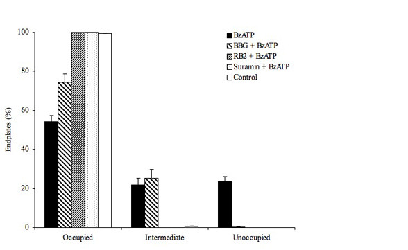
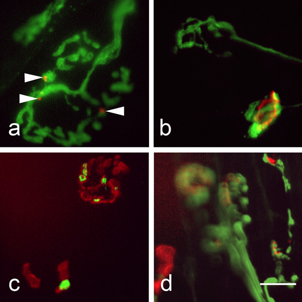
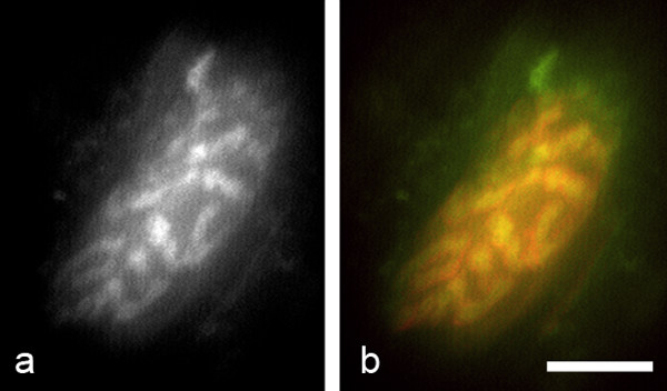
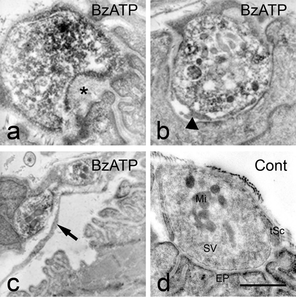
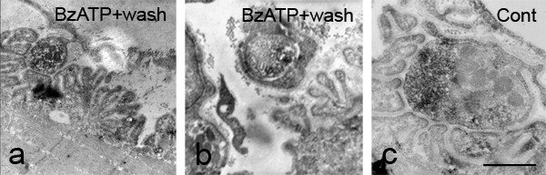
Results: We used an isolated preparation consisting of distal peripheral nerve stumps, associated nerve terminals and post-synaptic muscle fibres, maintained in-vitro for up to 3 hrs. In this system synapses are intact but the presynaptic nerve terminal is disconnected from its cell soma. In control preparations synapses were stable for extended periods and did not undergo Wallerian degeneration. In contrast, addition of purines triggers rapid changes at synapses. Using fluorescence and electron microscopy we observe ultrastructural and gross morphological events consistent with nerve terminal retraction. We find no evidence of Wallerian or Wallerian-like degeneration in these preparations. Pharmacological experiments implicate pre-synaptic P2X7 receptor subunits as key mediators of these events.

Conclusion: The data presented suggest; first that isolated nerve terminals are able to regulate connectivity independent of signals from the cell body, second that synapses exist in a dynamic state, poised to shift from stability to loss by activating intrinsic mechanisms and molecules, and third that local purines acting at purinergic receptors can trigger these events. A role for ATP receptors in this is not surprising since they are frequently activated during cellular injury, when adenosine tri-phosphate is released from damaged cells. Local control demands that the elements necessary to drive retraction are constitutively present. We hypothesize that pre-existing scaffolds of molecular motors and cytoskeletal proteins could provide the dynamism required to drive such structural changes in nerve terminals in the absence of the cell body.


Figures

Figure 1
Figure 1
Nerve terminal bouton loss in response to an extracellular cue occurs in the absence of neuronal cell bodies. All preparations were fixed in 4% paraformaldehyde prior to immunostaining for neurofilament 165 and SV2 (green: axons and terminals), visualised with a Cy2-conjugated secondary antibody and co-stained with TRITC-αBTX (red: muscle endplates), 165 min after a 15 min BzATP pulse (100 μM). Both 'occupied' (a: arrow) and 'unoccupied' (a: arrowhead) endplates were present, as were unoccupied endplates with intact presynaptic axons (b). Many terminals appeared to be in the process of retraction resulting in minor (c: asterisk) or major (d: asterisks) regions of unoccupied endplates. These were classified as 'intermediate' in subsequent data. Control nerve/muscle preparations, which were maintained in physiological saline for up to 300 min, showed no visible signs of degeneration or retraction of nerve terminals (e). Scale bar a-e = 15 μm
Figure 2
Figure 2
Nerve terminal loss appears to be triggered via activation of purinoceptors. Figure showing the proportion of occupied, intermediate and unoccupied muscle endplates present in: 300 min control (open bar); 165 min subsequent to a 15 min BzATP pulse (100 μM: filled bar); 165 min subsequent to a 15 min BzATP pulse (filled bar) in the presence of Brilliant Blue G (a selective P2X7 receptor antagonist: BBG, 1 μM: broad diagonal cross-hatching); 165 min subsequent to a 15 min BzATP pulse in the presence of Reactive Blue 2 (which blocks P2Y and P2X receptor subunits: 100 μM, RB2: narrow diagonal cross-hatching) or 165 min subsequent to a 15 min BzATP pulse in the presence of Suramin (a broad spectrum P2 receptor agonist: 100 μM: stipple). All data is mean ± SEM of multiple repeats (see text for n values).
Figure 3
Figure 3
Some nerve terminals and muscle endplates fragmented in response to BzATP. All preparations were fixed in 4% paraformaldehyde prior to immunostaining for neurofilament 165 and SV2 (green: axons and terminals), visualised with a Cy2-conjugated secondary antibody and co-stained with TRITC-αBTX (red: muscle endplates), 165 min after a 15 min BzATP (100 μM) pulse. Nerve terminals associated with either fragmented (a), or absent (b) muscle endplates were seen, as were fragmented nerve terminals contacting either apparently normal (c), or fragmented endplates (c, d). Scale bar a, d = 15 μm, b, c = 30 μm
Figure 4
Figure 4
Intact nerve terminals were capable of releasing and recycling synaptic vesicles subsequent to the addition of BzATP. The fluorescent vital styryl dye RH414 was used to label recycling synaptic vesicles by indirect nerve stimulation at the end of the 180 min experimental period and just prior to fixation. Remaining nerve terminals were brightly labelled with the dye (a) and fully occupied underlying motor endplates (b). A grayscale image of a single nerve terminal is shown in panel a, while the same terminal (coloured green) is seen to fully occupy a singe motor endplate (coloured red) in the composite image. Scale bar = 10 μm
Figure 5
Figure 5
Ultrastructural events indicate that nerve terminal loss progresses by retraction. Nerve and muscle preparations were fixed for immuno-electron microscopy and labelled for P2X7 receptor subunits (dark DAB reaction product) to facilitate localisation. When preparations were fixed and immunostained subsequent to a 30 min BzATP (100 μM) pulse frequent abnormal appearances were found. These included; widened synaptic clefts with the appearance of mega-omega profiles (asterisk) (a), fine processes of terminal Schwann cells invaded the synaptic cleft (b: arrowhead), boutons which were detached from the muscle endplate and where terminal Schwann cell processes had come to intervene between the two (c: arrow). In control nerve muscle preparations incubated in physiological saline for 30 min prior to fixation and immunostaining (d), motor nerve terminal boutons contained reaction product for P2X7RS, intact mitochondria (Mi), densely packed synaptic vesicles (SV), were closely apposed to muscle endplates (EP) and overlain by terminal Schwann cell (tSc). a, b, d = 500 nM, c = 1 μM
Figure 6
Figure 6
Ultrastructural changes compatible with nerve terminal retraction continue after removal of BzATP. When nerve/muscle preparations were exposed to a 30 min BzATP (100 μM) pulse followed by a 60 min washout period prior to fixation, many nerve terminal boutons were in advanced states of retraction. This was manifested as vacant regions of muscle endplate (a) and vesicle-filled boutons that were clearly wrapped by Schwann cell processes (b). Control preparations maintained in physiological saline for 90 min (c) were identical in appearance to freshly fixed preparations (compare with figure 5a). Scale bar a, b = 1 μM, c = 500 nM

Similar articles

Cited by

References

    1. Lichtman JW, Colman H. Synapse elimination and indelible memory. Neuron. 2000;25:269–278. doi: 10.1016/S0896-6273(00)80893-4. - DOI - PubMed
    1. Lohof AM, Delhayebouchaud N, Mariani J. Synapse Elimination In the Central-Nervous-System – Functional- Significance and Cellular Mechanisms. Rev Neurosci. 1996;7:85–101. - PubMed
    1. Walsh MK, Lichtman JW. In vivo time-lapse imaging of synaptic takeover associated with naturally occurring synapse elimination. Neuron. 2003;37:67–73. doi: 10.1016/S0896-6273(02)01142-X. - DOI - PubMed
    1. Keller-Peck CR, Walsh MK, Gan WB, Feng G, Sanes JR, Lichtman JW. Asynchronous synapse elimination in neonatal motor units: studies using GFP transgenic mice. Neuron. 2001;31:381–394. doi: 10.1016/S0896-6273(01)00383-X. - DOI - PubMed
    1. Costanzo EM, Barry JA, Ribchester RR. Competition at silent synapses in reinnervated skeletal muscle. Nat Neurosci. 2000;3:694–700. doi: 10.1038/76649. - DOI - PubMed

Publication types