. 2008 Apr;197(2):534-40.
doi: 10.1016/j.atherosclerosis.2007.08.015. Epub 2007 Sep 27.

Absence of regulated splicing of fibronectin EDA exon reduces atherosclerosis in mice

Affiliations

Absence of regulated splicing of fibronectin EDA exon reduces atherosclerosis in mice

Vladimir R Babaev et al. Atherosclerosis. 2008 Apr.

Abstract

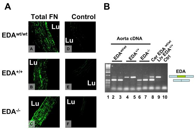
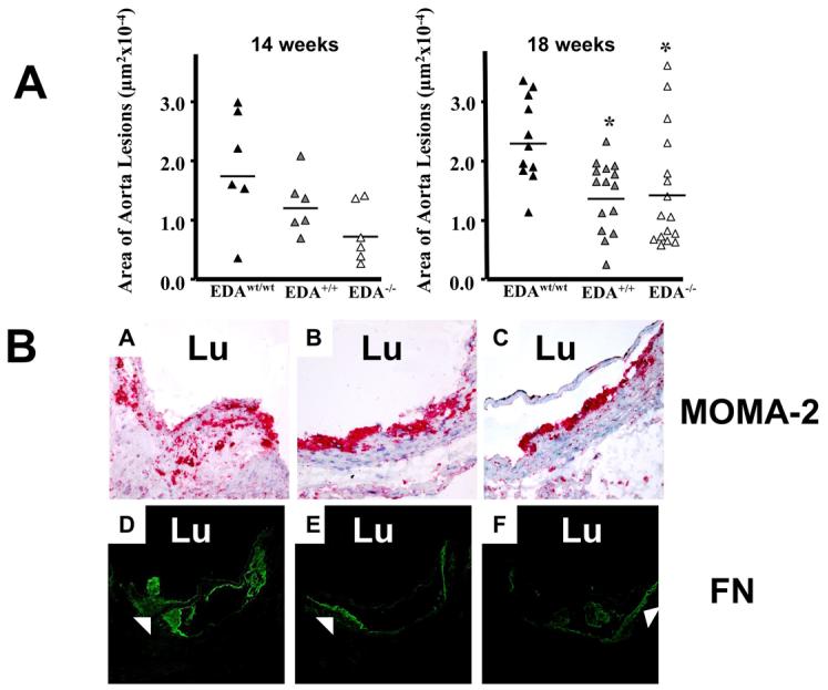
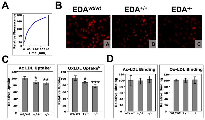
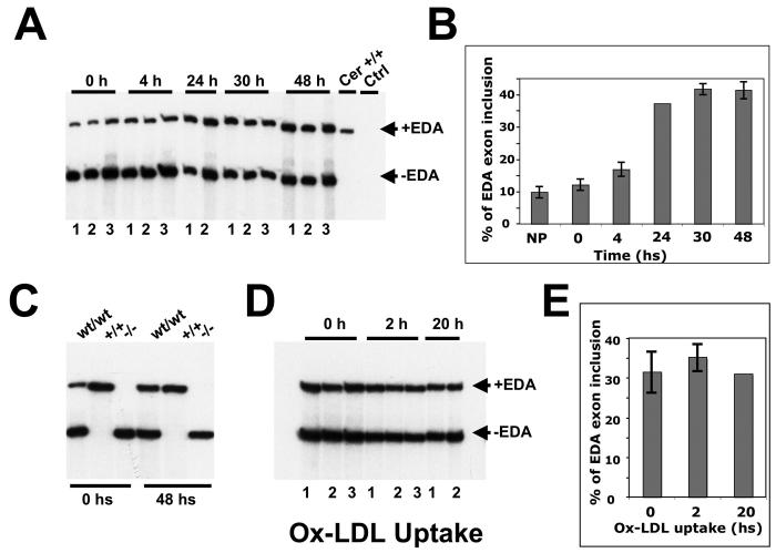
Atherosclerotic lesions are characterized by a profound alteration in the architecture of the arterial intima, with a marked increase of fibronectin (FN) and the appearance of the alternatively spliced FN variant containing the extra domain A (EDA). To analyze the role of FN isoforms in atherosclerotic lesion formation we utilized mouse strains devoid of EDA exon regulated splicing, which constitutively include (EDA(+/+)) or exclude (EDA(-/-)) the exon. Both mutant mice had a 40% reduction in atherosclerotic lesions after the atherogenic-diet treatment (mean+/-S.E., microm(2); 22969+/-2185; 13660+/-1533; 14260+/-2501 for EDA(wt/wt), EDA(+/+) and EDA(-/-), respectively; p< or =0.01 ANOVA test) associated to a lower capacity of macrophages to uptake modified LDL and undergo foam-cell formation. Lesions in control mice were more numerous and bigger, with augmented and deeper macrophage infiltration, and increased FN expression in the sub-endothelial area. Previous experiments have shown that apoE(-/-)EDA(-/-) mice have a decreased number and size of atherosclerotic lesions and, on this basis, it has been proposed that the EDA domain has a pro-atherogenic role. Our data with the EDA(+/+) mice rules out this hypothesis and suggest that regulated splicing of the EDA exon of the FN gene is involved in progression of atherosclerosis, highlighting the importance of alternative splicing in regulating cellular processes.


Figures

Figure 1
Figure 1. Panel A. Immunohistochemical analysis of FN distribution in mouse aorta
Sections from the proximal aorta of EDAwt/wt (A and D), EDA+/+ (B and E) and EDA−/− mice (C and F) were incubated with anti-total FN rabbit polyclonal antibody (A, B, and C) or control antibody (D, E and F). Original magnification 650x; Panel B. RT-PCR analysis of FN-EDA expression in aorta samples. RT-PCR analysis of RNA prepared from total aorta of EDAwt/wt untreated 2-3 month old mice showed that FN is mainly of the EDA-minus form (Lanes 2-3). The EDA+/+ and EDA−/− mice showed complete inclusion and complete exclusion of the EDA exon, respectively (lanes 4-5 and 6-7, respectively). Cerebellum EDAwt/wt (Cer) and liver EDA+/+ are shown as controls (lanes 8 and 9, respectively). Lane 10 corresponds to PCR control without cDNA. “L” indicates vessel lumen.
Figure 2
Figure 2. Panel A. EDA+/+ and EDA−/− mice are protected against atherosclerosis after atherogenic-diet
The atherosclerotic lesion area in the proximal aorta region was analyzed at 14 and 18 weeks after the initiation of the diet. Each triangle corresponds to the mean values of 15 sections from a single mouse. Compared to the EDAwt/wt mice, the EDA+/+ and EDA−/− mice had lower atherosclerotic lesion area at both 14 and 18 weeks of diet (Mean±SE, μm2; 14 weeks: 17482±3716; 12028±2011; 7216±2036; 18 weeks: 22969±2185; 13660±1533; 14260±2501 for EDAwt/wt, EDA+/+ and EDA−/−, respectively; 14 weeks, n=6 mice per genotype; 18 weeks, n=11, 15 and 16 mice for EDAwt/wt, EDA+/+ and EDA−/− , respectively). Data was analyzed by ANOVA test (14 weeks, NS; 18 weeks, p≤0.01) with the Bonferroni's multiple comparison Test (18 weeks, EDAwt/wt vs. EDA+/+ p≤0.05; EDAwt/wt vs. EDA−/−, p≤0.05). Panel B. Detection of macrophages and the FN distribution in atherosclerotic lesions of the proximal aorta. Sections from the proximal aorta of EDAwt/wt (A and D), EDA+/+ (B and E) and EDA−/− mice (C and F) fed with atherogenic-diet for 18 weeks were stained with the MOMA-2 monoclonal antibody (A, B and C) or with the anti-FN polyclonal antibody (D, E and F). (Original magnification, 400x and 200x for MOMA-2 and FN antibodies, respectively). “Lu” indicates vessel lumen.
Figure 3
Figure 3. Panel A. Time course of the modified LDL uptake by EDAwt/wt peritoneal macrophages
Peritoneal macrophages were incubated with 15 μg/ml of human DiL-oxidized for the indicated times and the uptake was examined by flow cytometry. The data are presented in relative units as an average of triplicate repeats. Panel B. Visualization of the modified-LDL uptake by peritoneal macrophages from EDAwt/wt, EDA+/+ and EDA−/− mice. Peritoneal macrophages from EDAwt/wt (A), EDA+/+ (B) and EDA−/− mice (C) were incubated with 15 μg of Ac-DiL LDL for 30 min. and visualized under UV light. Original magnification, 400x; Panels C and D. FACS analysis of the modified-LDL uptake (C) and binding (D) by peritoneal macrophages from EDAwt/wt, EDA+/+ and EDA−/− mice. Peritoneal macrophages were incubated for 30 min at 37°C (uptake) or 60 min at 4°C (binding) with Ac-DiL LDL or Ox-DiL LDL and uptake was examined by flow cytometry. The data of the uptake are presented in relative units as an average of three independent experiments (each experiment having three mice per genotype, each mouse having three repeats) ± Standard Deviation. The data of the binding experiment are presented in relative units as an average of three mice per genotype, each mouse having three repeats (a, ANOVA p=0.0023; b, ANOVA, p=0.0002; Bonferroni's Correction: *, p≤0.05; **, p≤0.01; ***, p≤0.001). wt, +/+ and −/− indicate EDAwt/wt, EDA+/+ and EDA−/− samples, respectively.
Figure 4
Figure 4. RT-PCR analysis of the FN-EDA splicing of peritoneal macrophages from EDAwt/wt after incubation with modified Ox-DiL LDL
(A) Macrophages from three EDAwt/wt animals (numbered 1 to 3) were plated, washed after 40 minutes (t=0), total RNA was prepared at different times (t=4, 24, 30 and 48 hs) and analyzed by radioactive RT-PCR. Total RNA from EDA+/+ cerebellum was run as control. (B) The PCR products from Panel A were quantified using a Phosphor Imager. (C) Similarly, EDAwt/wt, EDA+/+ and EDA−/− samples were plated for 0 and 48 hs, the RNA prepared and analyzed by radioactive RT-PCR. (D) Macrophages from EDAwt/wt mice were prepared, plated and after 48 hs were incubated with Ox-DiL LDL for 2 and 20 additional hs. (E) The radioactivity was quantified. Each bar corresponds to macrophages prepared from separate EDAwt/wt mice (numbered 1 to 3). The data was analyzed by One Way ANOVA and is presented as the mean ± standard deviation. No differences were observed.

References

    1. Osterud B, Bjorklid E. Role of monocytes in atherogenesis. Physiol Rev. 2003;83:1069–112. - PubMed
    1. ffrench-Constant C. Alternative splicing of fibronectin--many different proteins but few different functions. Exp Cell Res. 1995;221:261–71. - PubMed
    1. Hynes RO. Fibronectins. Springer-Verlag; New York: 1990. p. 544.
    1. Kornblihtt AR, Pesce CG, Alonso CR, Cramer P, Srebrow A, Werbajh S, Muro AF. The fibronectin gene as a model for splicing and transcription studies. Faseb J. 1996;10:248–57. - PubMed
    1. Mosher DF. Fibronectin. Academic Press; New york: 1989. p. 474.

Publication types