

Review
doi: 10.1016/j.semcdb.2007.08.003.
Epub 2007 Aug 19.
Ras nanoclusters: molecular structure and assembly
Affiliations
- PMID: 17897845
- PMCID: PMC2761225
- DOI: 10.1016/j.semcdb.2007.08.003
Item in Clipboard
Review
Ras nanoclusters: molecular structure and assembly
Semin Cell Dev Biol.
2007 Oct.
Abstract
H-, N- and K-ras4B are lipid-anchored, peripheral membrane guanine nucleotide binding proteins. Recent work has shown that Ras proteins are laterally segregated into non-overlapping, dynamic domains of the plasma membrane called nanoclusters. This lateral segregation is important to specify Ras interactions with membrane-associated proteins, effectors and scaffolding proteins and is critical for Ras signal transduction. Here we review biological, in vitro and structural data that provide insight into the molecular basis of how palmitoylated Ras proteins are anchored to the plasma membrane. We explore possible mechanisms for how the interactions of H-ras with a lipid bilayer may drive nanocluster formation.
Figures
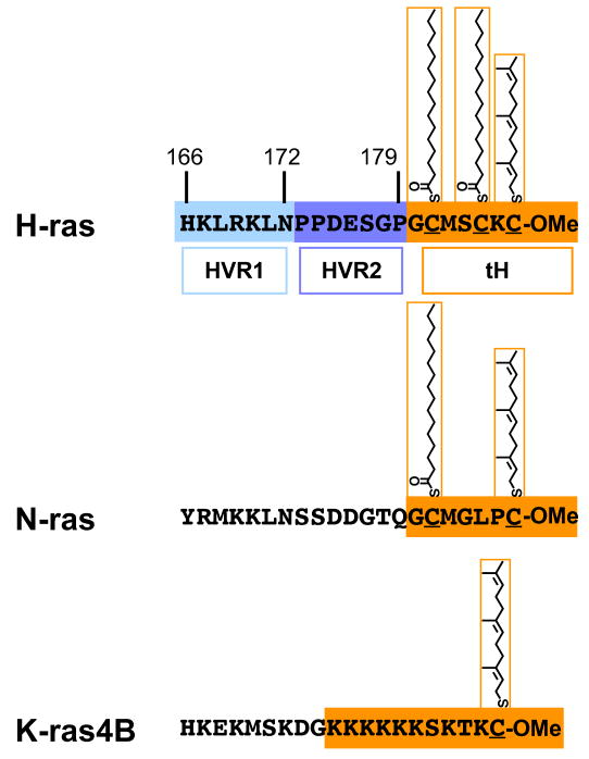
The processed hypervariable regions of H-, N- and K-ras. In the case of H-ras, the division of the linker region of the HVR into HVR1 and 2 (blue hues) is shown. The minimal plasma membrane targeting sequences are outlined in orange and include C-terminal farnesylation and carboxylmethylation, and the palmitoyl modifications or the stretch of basic residues. The minimal plasma membrane targeting sequence of H-ras is abbreviated tH.
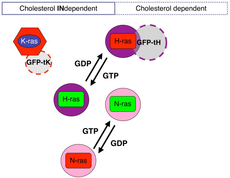
Nanodomains of the classical Ras isoforms characterized by EM and statistical analysis of the point patterns of immunogold-labelled plasma membrane sheets. Thus, both the sensitivity of nanoclustering towards cholesterol depletion or the co-clustering of two proteins can be examined. Both H-ras and N-ras show GTP-dependent lateral segregation into nanoclusters with different sensitivities to cholesterol depletion. It is unclear whether K-ras exhibits a similar GTP-dependent lateral segregation. Note that only proteins in nanoclusters are shown; randomly distributed proteins are omitted.
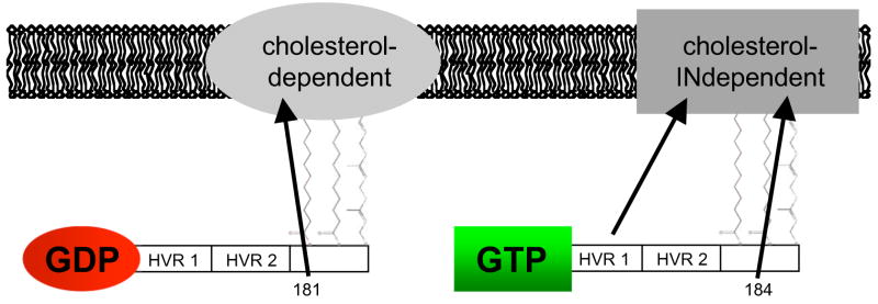
Vectorial model of GTP-dependent H-ras lateral segregation as defined by EM and FRAP analysis. This model complements a previous vectorial model in Rotblat et al. (2004) [36], by adding the contributions from the individual palmitoyls and HVR1.
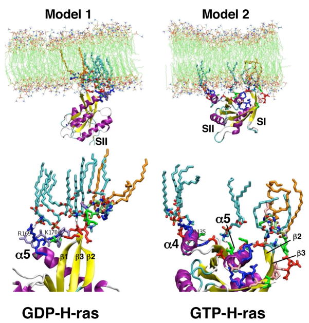
The two models of membrane bound H-ras obtained from MD simulations. Model 1(left) is the predominant conformation found for H-ras-GDP, while model 2 (right) is the predominant conformation for H-ras-GTP. The blowup of the interaction sites (bottom) shows lipids (cyan) and interacting residues of H-ras in sticks. Note that model 1 is predominantly anchored via the HVR, while model 2 shows extensive contact of α-helix 4 with the membrane. Membrane lipids (cyan) with specific contacts to H-ras residues or its lipids (orange) are shown in sticks.

Summary of mechanisms by which membrane insertion of Ras could drive nanoclustering. Differences in membrane insertion of GDP- and GTP-loaded H-ras provide some intriguing insights in the biophysical principals that may drive activation-dependent lateral segregation.
References
-
- Hancock JF, Magee AI, Childs JE, Marshall CJ. Cell. 1989;57:1167–77. - PubMed
-
- Bergo MO, Lieu HD, Gavino BJ, Ambroziak P, Otto JC, Casey PJ, Walker QM, Young SG. J Biol Chem. 2004;279:4729–36. - PubMed
-
- Pillinger MH, Volker C, Stock JB, Weissmann G, Philips MR. J Biol Chem. 1994;269:1486–92. - PubMed
-
- Hancock JF. Nat Rev Mol Cell Biol. 2003;4:373–84. - PubMed
Publication types
MeSH terms
Substances
Grants and funding
LinkOut - more resources
Full Text Sources
Other Literature Sources
Research Materials
Miscellaneous
