. 2007;35(19):6526-38.
doi: 10.1093/nar/gkm727. Epub 2007 Sep 26.

Multiple conserved regulatory elements with overlapping functions determine Sox10 expression in mouse embryogenesis

Affiliations

Multiple conserved regulatory elements with overlapping functions determine Sox10 expression in mouse embryogenesis

Torsten Werner et al. Nucleic Acids Res. 2007.

Abstract

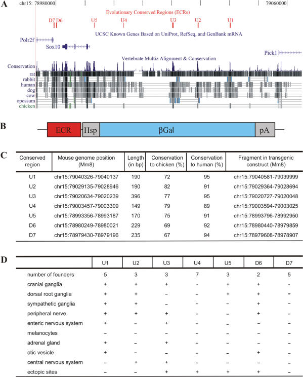
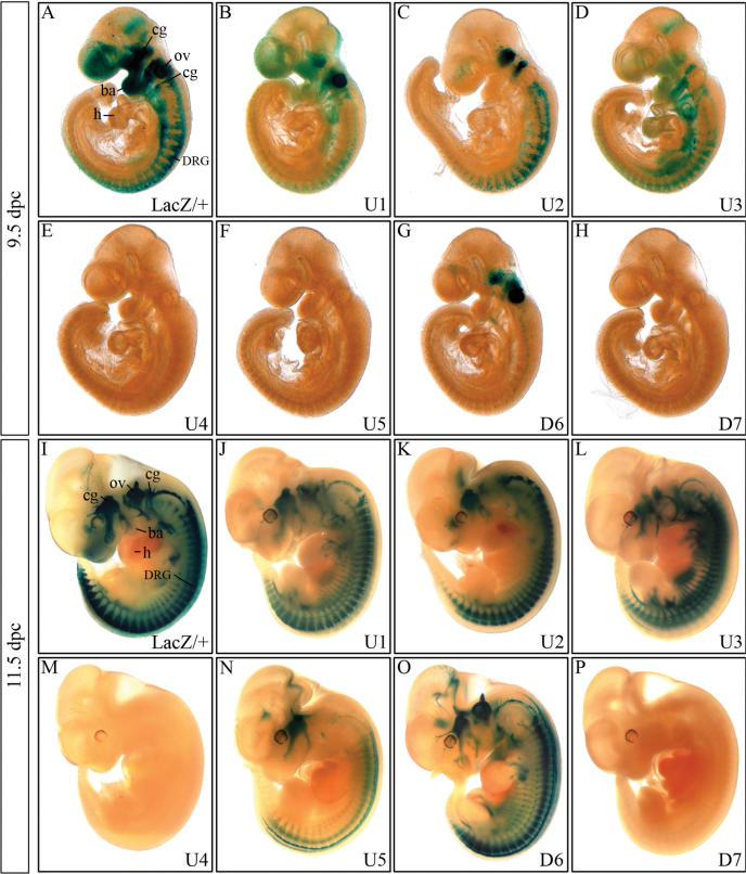
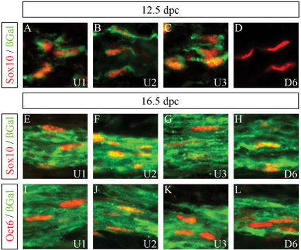
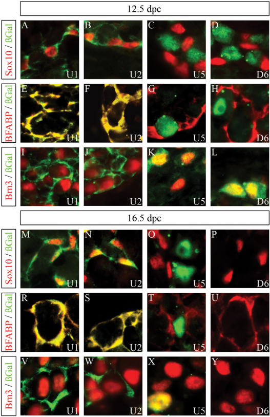
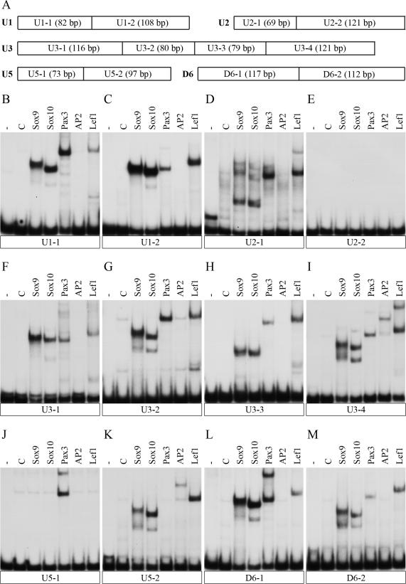
Expression and function of the transcription factor Sox10 is predominant in neural crest cells, its derivatives and in oligodendrocytes. To understand how Sox10 expression is regulated during development, we analysed the potential of evolutionary conserved non-coding sequences in the Sox10 genomic region to function as enhancers. By linking these sequences to a beta-galactosidase marker gene under the control of a minimal promoter, five regulatory regions were identified that direct marker gene expression in transgenic mice to Sox10 expressing cell types and tissues in a defined temporal pattern. These possible enhancers of the Sox10 gene mediate Sox10 expression in the otic vesicle, in oligodendrocytes and in several neural crest derivatives including the developing peripheral nervous system and the adrenal gland. They furthermore exhibit overlapping activities and share binding sites for Sox, Lef/Tcf, Pax and AP2 transcription factors. This may explain high level and robustness of Sox10 expression during embryonic development.


Figures

Figure 1.
Figure 1.
Characterization of evolutionary conserved non-coding sequences from the Sox10 genomic region and generation of transgenic lines containing these sequences. (A) Localization of seven ECR (U1, U2, U3, U4, U5, D6, D7, shown in red) in the Sox10 genomic interval on mouse chromosome 15 relative to Sox10 and the adjacent Polr2f and Pick1 genes (in blue). Sequence conservation among various mammalian species and chicken is evident from multiple sequence alignments. (B) Design of the transgenic constructs consisting of one of the seven ECR, the hsp68 minimal promoter (Hsp), the β-galactosidase marker gene (βGal) and a SV40 polyA signal (pA). (C) Position of each of the seven ECR in the mouse genome (Mm8), their lengths, conservation to chicken and man as well as the exact coordinates of the fragment present in the transgenic constructs. (D) Summary of independent founders obtained for each transgenic construct, resulting transgenic lines and their overall properties of transgene expression.
Figure 2.
Figure 2.
Detection of β-galactosidase transgene expression in embryos at 9.5 and 11.5 d.p.c. β-Galactosidase activity was detected colorimetrically using X-gal substrate at 9.5 dpc (AH) and at 11.5 d.p.c. (IP) in Sox10lacZ/+ (A, I) and age-matched embryos carrying a hsp68-lacZ transgene driven by one of the following ECR from the Sox10 genomic region: U1 (B, J), U2 (C, K), U3 (D, L), U4 (E, M), U5 (F, N), D6 (G, O) and D7 (H, P). Transgenic embryos were stained in parallel for 9 h, Sox10lacZ/+ embryos for 3 h. No β-galactosidase staining was detected in wildtype littermates under the conditions applied. cg, cranial ganglia; DRG, dorsal root ganglia; ov, otic vesicle; ba, branchial arches; h, heart.
Figure 3.
Figure 3.
Cellular expression pattern of β-galactosidase transgenes in peripheral nerves. Co-immunohistochemistry was performed on spinal nerves of embryos at 12.5 d.p.c. (AD) and 16.5 d.p.c. (EL) using antibodies directed against β-galactosidase (in green) in combination with antibodies directed against Sox10 (A–H) or Oct6 (I–L) (all in red). The embryos carried a hsp68-lacZ transgene driven by either the U1 (A, E, I), the U2 (B, F, J), the U3 (C, G, K) or the D6 (D, H, L) region.
Figure 4.
Figure 4.
Cellular expression pattern of β-galactosidase transgenes in DRG. Co-immunohistochemistry was performed on DRG of embryos (forelimb level) at 12.5 d.p.c. (AL) and 16.5 d.p.c. (MY) using antibodies directed against β-galactosidase (in green) in combination with antibodies directed against Sox10 (A–D, M–P), BFABP (E–H, R–U) or Brn3.0 (I–L, V–Y) (all in red). The embryos carried a hsp68-lacZ transgene driven by either the U1 (A, E, I, M, R, V), the U2 (B, F, J, N, S, W), the U5 (C, G, K, O, T, X) or the D6 (D, H, L, P, U, Y) region.
Figure 5.
Figure 5.
Detection of β-galactosidase transgene expression in embryos at 16.5 d.p.c. β-Galactosidase activity was detected colorimetrically using X-gal substrate on transverse sections from the forelimb level of Sox10lacZ/+ (A) and age-matched embryos carrying a hsp68-lacZ transgene driven by one of the following ECR from the Sox10 genomic region: U1 (B), U2 (C), U3 (D), U5 (E) and D6 (F). Embryo sections were stained in parallel for 2 h. No β-galactosidase staining was detected in wildtype littermates under the conditions applied.
Figure 6.
Figure 6.
Cellular expression pattern of β-galactosidase transgenes in the embryonic spinal cord. Co-immunohistochemistry was performed on transverse sections of the embryonic spinal cord (forelimb level) at 16.5 d.p.c. using antibodies directed against β-galactosidase (in green) in combination with antibodies directed against the cell-type specific markers (in red) Sox10 (A, C), Olig2 (B) and Lmx1 (D). The embryos carried a hsp68-lacZ transgene driven by the U2 (A, B) or the U3 (C, D) region.
Figure 7.
Figure 7.
Binding of neural crest derived transcription factors to evolutionary conserved non-coding sequences from the Sox10 genomic region. (A) Schematic representation of the fragments used for EMSA. The length of each fragment is given in bp. (BM) Each of the radiolabeled fragments shown in (A) was incubated with control extracts (C) or extracts containing Sox9, Sox10, Pax3, AP2α or Lef1 protein before protein-DNA complexes were resolved from the unbound DNA by native gel electrophoresis. The unbound DNA is shown in the first lane of each panel (-) and runs at the bottom. Each panel represents the results from EMSA of a single fragment: (B) U1-1; (C) U1-2; (D) U2-1; (E) U2-2; (F) U3-1; (G) U3-2; (H) U3-3; (I) U3-4; (J) U5-1); (K) U5-2; (L) D6-1; (M) D6-2.

References

    1. Aoki Y, Saint-Germain N, Gyda M, Magner-Fink E, Lee YH, Credidio C, Saint-Jeannet JP. Sox10 regulates the development of neural crest-derived melanocytes in Xenopus. Dev. Biol. 2003;259:19–33. - PubMed
    1. Cheung M, Briscoe J. Neural crest development is regulated by the transcription factor Sox9. Development. 2003;130:5681–5693. - PubMed
    1. Dutton KA, Pauliny A, Lopes SS, Elworthy S, Carney TJ, Rauch J, Geisler R, Haffter P, Kelsh RN. Zebrafish colourless encodes sox10 and specifies non-ectomesenchymal neural crest fates. Development. 2001;128:4113–4125. - PubMed
    1. Herbarth B, Pingault V, Bondurand N, Kuhlbrodt K, Hermans-Borgmeyer I, Puliti A, Lemort N, Goossens M, Wegner M. Mutation of the Sry-related Sox10 gene in Dominant megacolon, a mouse model for human Hirschsprung disease. Proc. Natl Acad. Sci. USA. 1998;95:5161–5165. - PMC - PubMed
    1. Honore SM, Aybar MJ, Mayor R. Sox10 is required for the early development of the prospective neural crest in Xenopus embryos. Dev. Biol. 2003;260:79–96. - PubMed

Publication types

MeSH terms