. 2007 Dec;81(24):13649-58.
doi: 10.1128/JVI.01385-07. Epub 2007 Sep 26.

Development of a challenge-protective vaccine concept by modification of the viral RNA-dependent RNA polymerase of canine distemper virus

Affiliations

Development of a challenge-protective vaccine concept by modification of the viral RNA-dependent RNA polymerase of canine distemper virus

D Silin et al. J Virol. 2007 Dec.

Abstract

We demonstrate that insertion of the open reading frame of enhanced green fluorescent protein (EGFP) into the coding sequence for the second hinge region of the viral L (large) protein (RNA-dependent RNA polymerase) attenuates a wild-type canine distemper virus. Moreover, we show that single intranasal immunization with this recombinant virus provides significant protection against challenge with the virulent parental virus. Protection against wild-type challenge was gained either after recovery of cellular immunity postimmunization or after development of neutralizing antibodies. Insertion of EGFP seems to result in overattenuation of the virus, while our previous experiments demonstrated that the insertion of an epitope tag into a similar position did not affect L protein function. Thus, a desirable level of attenuation could be reached by manipulating the length of the insert (in the second hinge region of the L protein), providing additional tools for optimization of controlled attenuation. This strategy for controlled attenuation may be useful for a "quick response" in vaccine development against well-known and "new" viral infections and could be combined efficiently with other strategies of vaccine development and delivery systems.


Figures
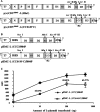
FIG. 1.
FIG. 1.
Construction of recombinant viruses. The positions of the T7 promoter (T7), the internal ribosome entry site (IRES), three domains of the RdRp (D1 to D3), the hinge regions of the RdRp (H1 and H2), the hepatitis delta ribozyme (δ), and the T7 terminator (Tφ) are indicated (not to scale). Bold lines indicate the positions of the intergenic trinucleotide spacers. (A) Schematic representation of rCDV5804P and rCDV5804P-L(CCegfpC) full-length genome plasmids. Three conservative domains and two hinge regions of the L protein are shown. The EGFP ORF was cloned into the MluI site. (B) Schematic representation of plasmids expressing rCDV5804P and rCDV5804P-L(CCegfpC) L proteins. (C) Relative activities of unmodified and EGFP-modified CDV5804P polymerases. Various amounts of pEMC-L (CCC)5804P (diamonds) and pEMC-L(CCegfpC)5804P (squares) were used in minigenome transfection assays. The amount of DsRed2 produced was determined by flow cytometry and expressed in relative fluorescence units (RFU). Percentages represent the relative activities of the modified RdRp compared to the unmodified form of the protein.
FIG. 2.
FIG. 2.
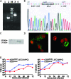
Rescue of recombinant viruses. (A) RT-PCR products generated with primers spanning the MluI site in the L protein gene. Lanes: 1, no-RT control with RNA extracted from CDV5804P-infected cells; 2, RNA extracted from CDV5804P-infected cells (926-bp product); M, DNA marker (λ DNA digested with PstI); 3, RNA extracted from rCDV5804P-L(CCegfpC)-infected cells (1,646-bp product); 4, no-RT control with RNA extracted from rCDV5804P-L(CCegfpC)-infected cells. (B) Sequence analysis of RT-PCR product from lane 4 in panel A indicating that the EGFP ORF is inserted in frame and in the correct orientation. (C) Western blot developed with polyclonal anti-GFP antibodies. SDS-PAGE was carried out under reducing conditions. Left lane, VeroDogSLAM cell lysate; right lane, lysate of VeroDogSLAM cells infected with rCDV5804P-L(CCegfpC). (D) Autofluorescence of rCDV5804P-L(CCegfpC)-infected VeroDogSLAM cells (left) and ferret lymphocytes (right). The nuclei were stained with propidium iodide, and the signal in the green channel was produced by autofluorescence of the L protein. (E) Growth curves of viruses on VeroDogSLAM cells. Cells were infected at an MOI of 0.01. Viral titers in the supernatant (left panel) and cell-associated viral titers (right panel) are shown.
FIG. 3.
FIG. 3.
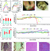
Disease development in animals infected with rCDV5804P only (A, C to F, and I to K) or infected with rCDV5804P-L(CCegfpC) (B) and then challenged with rCDV5804P (G and H). (A and B) Ferret body weight dynamics after primary infection. (C) Abdominal skin rash. (D) Mucopurulent nasal and ocular discharge. (E and G) Individual data on rash onset, recovery, and terminal stage of disease. (F and H) Individual body temperature curves postinfection (F) and postchallenge (H). †, ferrets were severely moribund and were euthanized. (I and J) Histopathology of lung tissue showing acute purulent bronchopneumonia. Hematoxylin-eosin staining (I) and immunohistochemical staining with monoclonal antibodies (VMRD, Inc.) against the CDV N protein (J) were performed. (K and L) Histology structure of spleens from animals infected with rCDV5804P (K) or infected with rCDV5804P-L(CCegfpC) and then challenged with rCDV5804P (L). Note the lymphoid depletion and absence of lymphoid follicles in the spleen of the moribund animal.
FIG. 4.
FIG. 4.
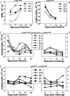
Hematological changes during infection. (A) WBC counts in peripheral blood. The curves show individual data for the group primarily infected with rCDV5804P. (B to F) Differential counts of lymphocytes. The curves show individual data for the group primarily infected with rCDV5804P (B) and the groups infected with rCDV5804P-L(CCegfpC) (C) and rCDVOP (E) and then challenged with rCDV5804P (D and F, respectively). Legends display animal numbers.
FIG. 5.
FIG. 5.
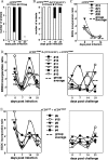
Nonspecific cellular immunity assessment. DTH reaction profiles of animals infected with rCDV5804P (A) or rCDV5804P-L(CCegfpC) (B) are shown. Proliferation potencies of PHA-stimulated PBMCs from individual animals in the group primarily infected with rCDV5804P (C) and in the groups infected with rCDV5804P-L(CCegfpC) (D) and rCDVOP (E) and then challenged with rCDV5804P are also shown. Legends display animal numbers. Group averages are shown by bold horizontal lines. BRDU, bromodeoxyuridine.
FIG. 6.
FIG. 6.
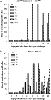
Development of specific anti-CDV neutralizing antibodies. Plaque neutralization titers for individual animals infected with rCDV5804P-L(CCegfpC) (A) and rCDVOP (B) followed by challenge with rCDV5804P are shown as reciprocal dilutions.

Similar articles

Cited by

References

    1. Appel, M. J., and B. A. Summers. 1995. Pathogenicity of morbilliviruses for terrestrial carnivores. Vet. Microbiol. 44:187-191. - PubMed
    1. Barrett, T. 1999. Morbillivirus infections, with special emphasis on morbilliviruses of carnivores. Vet. Microbiol. 69:3-13. - PubMed
    1. Bellini, W. J., G. Englund, S. Rozenblatt, H. Arnheiter, and C. D. Richardson. 1985. Measles virus P gene codes for two proteins. J. Virol. 53:908-919. - PMC - PubMed
    1. Brown, D. D., F. M. Collins, W. P. Duprex, M. D. Baron, T. Barrett, and B. K. Rima. 2005. ‘Rescue’ of mini-genomic constructs and viruses by combinations of morbillivirus N, P and L proteins. J. Gen. Virol. 86:1077-1081. - PubMed
    1. Brown, D. D., B. K. Rima, I. V. Allen, M. D. Baron, A. C. Banyard, T. Barrett, and W. P. Duprex. 2005. Rational attenuation of a morbillivirus by modulating the activity of the RNA-dependent RNA polymerase. J. Virol. 79:14330-14338. - PMC - PubMed

Publication types

MeSH terms

Substances

LinkOut - more resources