. 2007 Dec;81(23):13180-90.
doi: 10.1128/JVI.01400-07. Epub 2007 Sep 26.

Atraumatic oral spray immunization with replication-deficient viral vector vaccines

Affiliations

Atraumatic oral spray immunization with replication-deficient viral vector vaccines

Christiane Stahl-Hennig et al. J Virol. 2007 Dec.

Abstract

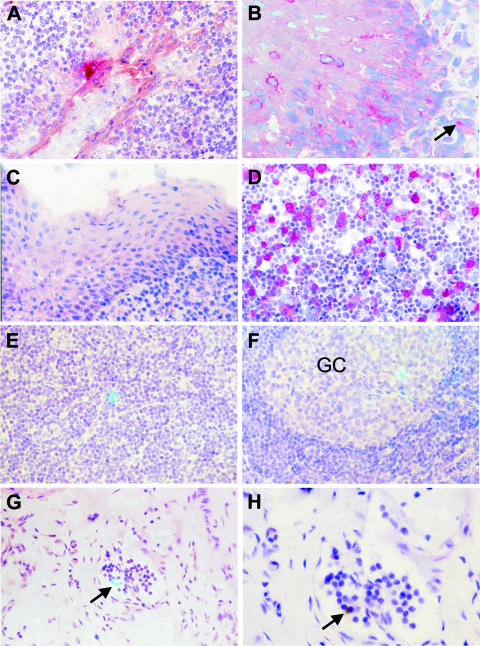
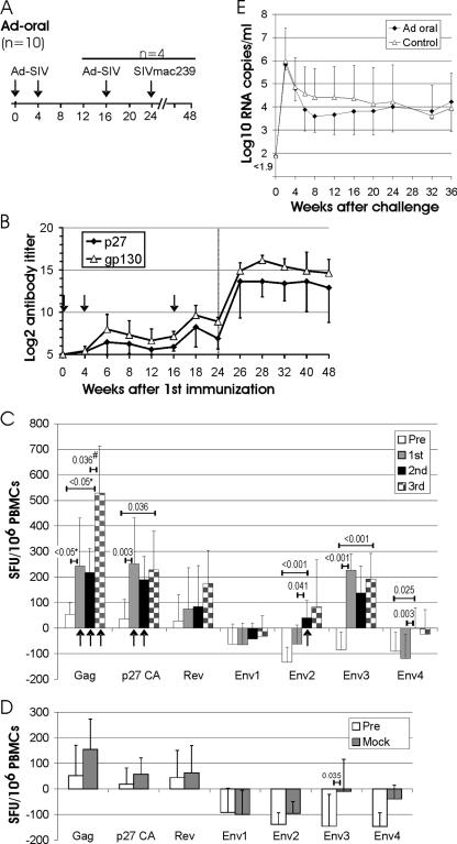
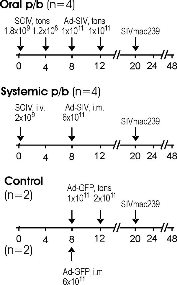
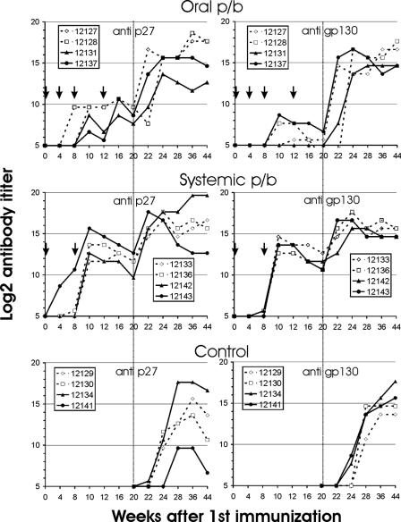
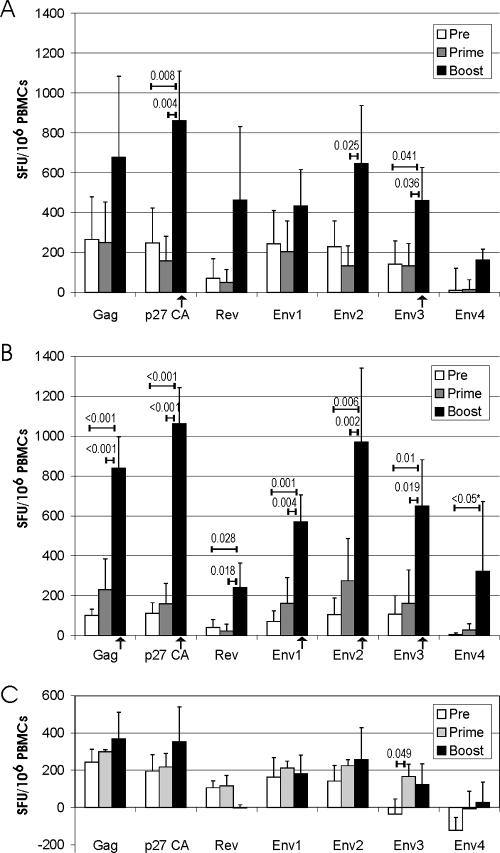
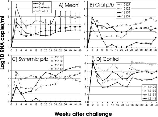
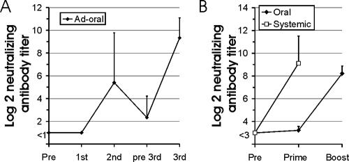
The development of needle-free vaccines is one of the recently defined "grand challenges in global health" (H. Varmus, R. Klausner, R. Klausner, R. Zerhouni, T. Acharya, A. S. Daar, and P. A. Singer, Science 302:398-399, 2003). To explore whether a natural pathway to the inductive site of the mucosa-associated lymphatic tissue could be exploited for atraumatic immunization purposes, replication-deficient viral vector vaccines were sprayed directly onto the tonsils of rhesus macaques. Tonsillar immunization with viral vector vaccines encoding simian immunodeficiency virus (SIV) antigens induced cellular and humoral immune responses. Viral RNA levels after a stringent SIV challenge were reduced, providing a level of protection similar to that observed after systemic immunization with the same vaccines. Thus, atraumatic oral spray immunization with replication-deficient vectors can overcome the epithelial barrier, deliver the vaccine antigen to the mucosa-associated lymphatic tissue, and avoid induction of tolerance, providing a novel approach to circumvent acceptability problems of syringe and needle vaccines for children and in developing countries.


Figures

FIG. 1.
FIG. 1.
Detection of replication-deficient viral vectors in the lymphoid tissue. (A and B) GFP expression after tonsillar administration of an adenoviral vector (Ad-GFP) was detected by immunohistochemistry (red) in the crypt (A) and squamous epithelium (B) of the tonsil. The arrow (B) points to a large, nonlymphoid cell showing positivity for GFP in the lymphoid tissue. (C) In a control macaque, tonsils were not stained with the antibody to GFP. (D) As a positive control, 293T cells transfected with a GFP expression plasmid were also stained. (E to H) SCIV-producing cells in axillary lymph nodes 4 days after intravenous inoculation (E) or tonsillar application (F to H) of the vaccine. Cells expressing SIV RNA (greenish blue color with the combined reflected light and transillumination) are in the T-dependent zone (E), the germinal center (GC), and the efferent lymphatic vessel (G and H). This vessel harboring the SCIV-positive cell (G, arrow) is shown with higher magnification (H). The morphology of the positive cell (H, arrow) demonstrates that it is a lymphocyte (transillumination only; the silver grains are black). The number of hybridization signals appears low since the silver grains of the signal are located in a higher focal plane than the cells of the tissue section. Original magnifications: A, C, and E to G, ×100; B and H, ×160; D, ×50.
FIG. 2.
FIG. 2.
Adenoviral vector immunization by the tonsillar route. (A) Experimental outline. The scale indicates the number of weeks after the first immunization. The type of vaccine or challenge virus is given above the time line. Four of the 10 monkeys of the Ad-oral group received a third oral immunization at week 16. (B) Mean titer and standard deviation of antibodies binding p27 CA or gp130 SU in 10 (weeks 0 to 12) and 4 (weeks 16 to 48) macaques of the Ad-oral group. The vertical dotted line in panel B indicates the time of challenge. (C) ELISPOT responses to the indicated peptide pools in the Ad-oral group were determined 4 and 0 weeks prior to immunization (Pre), and 2 and 4 weeks after the first, second, and third oral immunization with the adenoviral vector. ELISPOTs of unstimulated control cultures from each time point were subtracted, and the means for all available time points were used as single Pre, first, second, and third values for each animal. The means and standard deviations of the Pre, first, second, and third values for all animals after stimulation with the indicated peptide pools are shown. To determine whether there is a statistically significant difference between the means of the Pre, first, and second, ELISPOT values for each peptide, one-way ANOVA was used, followed by pairwise multiple comparison using the Tukey test. Numbers above the horizontal bars give the respective P values if a significant difference between two time points was obtained by ANOVA. Columns marked with an arrow indicate ELISPOT responses that are significantly higher (P < 0.05, t test) than the mock ELISPOT response to the respective peptide pool of the Ad-control group (D). For the 4 of the 10 animals that received a third oral immunization, a paired t test (#) was used to determine significant increases in ELISPOT responses between the second and third immunizations. (D) IFN-γ ELISPOT responses in six control animals were determined weeks −4, 0 (Pre), and 8 (mock) as described in for panel C. The t test was used to determine statistically significant differences between the Pre and mock ELISPOT responses for each peptide. *, due to failure of the equal variance test, ANOVA on ranks followed by pairwise comparison using Dunn's method was performed. (E) RNA load after challenge with SIVmac239. The mean and standard deviation of the viral RNA loads in the four animals receiving three oral adenoviral vector immunizations (Ad-oral) and in eight control monkeys infected in parallel are shown.
FIG. 3.
FIG. 3.
Prime-boost (p/b) immunization with viral vector vaccines. The different treatment groups and number of animals per group are given. The scale indicates the number of weeks after the first immunization. The type of vaccine or challenge virus, route of immunization, and dosages are given above the time line. tons, tonsillar; i.v., intravenous; i.m., intramuscular.
FIG. 4.
FIG. 4.
Antibody titers to SIV p27 CA (left panels) and gp130 SU (right panels) after oral or systemic immunization and in control monkeys. The five-digit numbers are monkey designations, and arrows mark the time points of immunization. The vertical dotted lines indicate the time of challenge. p/b, prime-boost.
FIG. 5.
FIG. 5.
IFN-γ ELISPOT response after prime-boost immunization by the oral (A) or systemic (B) route. ELISPOT responses to the indicated peptide pools were determined 1 and 3 weeks prior to immunization (Pre), at weeks 4 and 8 (prime), and at weeks 10 and 12 (boost). ELISPOT responses are presented as described in the legend to Fig. 2. Numbers above the horizontal bars give the respective P values if a significant difference between two time points was obtained by ANOVA. Columns marked with an arrow indicate that the ELISPOT response is significantly higher (P < 0.05, t test) than the ELISPOT response to the respective peptide pool of the control group at the same time point (C). *, due to failure of the normality test, ANOVA on ranks followed by pairwise comparison using Dunn's method was performed. PBMC, peripheral blood mononuclear cell.
FIG. 6.
FIG. 6.
RNA load after challenge. (A) The means and standard deviations for the three groups are shown. (B to D) The viral RNA loads in monkeys immunized orally (B) or systemically (C) and in the nonimmunized control group (D) are shown for each of the macaques. The five-digit numbers are monkey designations. p/b, prime-boost.
FIG. 7.
FIG. 7.
Neutralizing antibodies to the adenoviral vector. (A) Neutralizing antibody titers to the adenoviral vector before the first (Pre) and third (pre 3rd) and 2 weeks after the first, second, and third adenoviral vector applications (dose, 2 × 10¹¹ particles) by the tonsillar route (for the immunization regimen, see Fig. 2A). (B) Neutralizing antibody titers to the adenoviral vector were determined before (Pre) and 4 weeks after the first (prime) and second (boost) adenoviral vector applications by the tonsillar (oral; dose, 1 × 10¹¹) or intramuscular (systemic; dose, 6 × 10¹¹) route. The two control macaques receiving Ad-GFP either orally of systemically (for the immunization regimen, see Fig. 3A) were included in the analyses. The means and standard deviations for each group are shown.

References

    1. Banks, W. A., E. O. Freed, K. M. Wolf, S. M. Robinson, M. Franko, and V. B. Kumar. 2001. Transport of human immunodeficiency virus type 1 pseudoviruses across the blood-brain barrier: role of envelope proteins and adsorptive endocytosis. J. Virol. 75:4681-4691. - PMC - PubMed
    1. Barouch, D. H. 2006. Rational design of gene-based vaccines. J. Pathol. 208:283-289. - PubMed
    1. Barouch, D. H., M. G. Pau, J. H. Custers, W. Koudstaal, S. Kostense, M. J. Havenga, D. M. Truitt, S. M. Sumida, M. G. Kishko, J. C. Arthur, B. Korioth-Schmitz, M. H. Newberg, D. A. Gorgone, M. A. Lifton, D. L. Panicali, G. J. Nabel, N. L. Letvin, and J. Goudsmit. 2004. Immunogenicity of recombinant adenovirus serotype 35 vaccine in the presence of pre-existing anti-Ad5 immunity. J. Immunol. 172:6290-6297. - PubMed
    1. Betts, M. R., M. C. Nason, S. M. West, S. C. De Rosa, S. A. Migueles, J. Abraham, M. M. Lederman, J. M. Benito, P. A. Goepfert, M. Connors, M. Roederer, and R. A. Koup. 2006. HIV nonprogressors preferentially maintain highly functional HIV-specific CD8+ T cells. Blood 107:4781-4789. - PMC - PubMed
    1. Bomsel, M. 1997. Transcytosis of infectious human immunodeficiency virus across a tight human epithelial cell line barrier. Nat. Med. 3:42-47. - PubMed

Publication types

MeSH terms

LinkOut - more resources