. 2007 Dec;81(23):13271-6.
doi: 10.1128/JVI.01647-07. Epub 2007 Sep 26.

Crimean-Congo hemorrhagic fever virus glycoprotein processing by the endoprotease SKI-1/S1P is critical for virus infectivity

Affiliations

Crimean-Congo hemorrhagic fever virus glycoprotein processing by the endoprotease SKI-1/S1P is critical for virus infectivity

Eric Bergeron et al. J Virol. 2007 Dec.

Abstract

Crimean-Congo hemorrhagic fever virus (CCHFV) causes severe human disease. The CCHFV medium RNA encodes a polyprotein which is proteolytically processed to yield the glycoprotein precursors PreGn and PreGc, followed by structural glycoproteins Gn and Gc. Subtilisin kexin isozyme-1/site-1 protease (SKI-1/S1P) plays a central role in Gn processing. Here we show that CCHFV-infected cells deficient in SKI-1/S1P produce no infectious virus, although PreGn and PreGc accumulated normally in the Golgi apparatus, the site of virus assembly. Only nucleoprotein-containing particles which lacked virus glycoproteins (Gn/Gc or PreGn/PreGc) were secreted. Complementation of SKI-1/S1P-deficient cells with a SKI-1/S1P expression vector restored release of infectious virus (>10(6) PFU/ml), confirming that SKI-1/S1P processing is required for incorporation of viral glycoproteins. SKI-1/S1P may represent a promising antiviral target.


Figures

FIG. 1.
FIG. 1.
Schematic representation of CCHFV M-encoded polyprotein domains and proteolytic processing. (A) Signal peptide (SP), mucin-like, GP38, Gn, NSm, and Gc M polyprotein domains are highlighted. Potential transmembrane domains (yellow) and signal peptidase cleavage sites are indicated by black arrows. Antibodies used in this study are indicated (with their binding regions in parentheses): 7F5 (PreGn/GP38), Gn ectodomain (Gn ect.) (Gn), and 11E7 (PreGc/Gc). Defined furin-like (RSKR247), SKI-1/S1P (RRLL519), and SKI-1/S1P-like (RKPL1040) cleavage sites are illustrated by inverted triangles. (B) The first proteolytic products are expected to occur cotranslationally or rapidly after the synthesis of the polyprotein. PreGn (140 kDa), NSm (15 kDa), and PreGc (85 kDa) are the results of these initial processing events. SKI-1/S1P and PreGc convertase will then cleave (indicated by arrows) PreGn and PreGc in the early secretory pathway. The PreGc cleavage also occurs early in the secretory pathway but the cognate protease (?) remains unidentified. (C) The activity of SKI-1/S1P and the PreGc convertase generates a nonstructural mucin-like GP38 protein of either 160 or 85 kDa, and the structural glycoproteins Gn (37 kDa) and Gc (75 kDa). (D) The mucin-like GP38 is further cleaved (arrow) by a furin-like protease in the late secretory pathway. (E) Furin-like enzyme cleavage results in production of a GP38 glycoprotein (38 kDa) and a mucin-like protein of unknown mass (? kDa).
FIG. 2.
FIG. 2.
CCHFV growth in the absence (−) of SKI-1/S1P or S2P. SRD12B cells (top, SKI-1/S1P−) or M19 cells (bottom, S2P−) were infected with CCHFV strain IbAr10200 at a multiplicity of infection of 0.5. Cells were fixed with 4% paraformaldehyde at the indicated times postinfection and irradiated with 2 × 10⁶ rads. Fixed cells were permeabilized with 0.5% Triton X-100 and processed by IFA with CCHFV hyperimmune mouse ascitic fluid developed against CCHFV IbAr10200, as described earlier (22), and anti-mouse Alexa 594 antibody (Invitrogen). Images were acquired with a 20× objective.
FIG. 3.
FIG. 3.
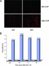
Production of infectious CCHFV from SKI-1/S1P-deficient cells can be rescued by plasmid-expressed SKI-1/S1P. (A) SKI-1/S1P-deficient cells (SRD12B cells) were transiently transfected with control empty vector (top, −SKI-1/S1P) or with SKI-1/S1P-containing vector (bottom, +SKI-1/S1P) by use of a Nucleofector II electroporator, the H-014 program, and cell line Nucleofector solution T (Amaxa, Gaithersburg, MD). These transfected cells were infected 24 hpi and processed at the indicated times postinfection as described in the legend for Fig. 2. No obvious cytopathic effect was observed in the presence or absence of SKI-1/S1P (data not shown). (B) Infections were set up as described above. Infected cell supernatants were collected, clarified, and frozen in liquid nitrogen at the indicated times for subsequent titration by plaque assay on SW13 cell monolayers (22). Red bars indicate viral titers from −SKI-1/S1P-transfected and blue bars from +SKI-1/S1P-transfected cell supernatants.
FIG. 4.
FIG. 4.
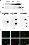
Glycoprotein-deficient viral particles are released in the infected cell supernatant of SKI-1/S1P-deficient cells. SRD12B cells stably expressing SKI-1/S1P were produced by transfection of the pIR-SKI-1/S1P vector with Lipofectamine 2000. Two days after transfection, cells were placed in Dulbecco's modified Eagle's medium-F-12 medium supplemented with 5% lipoprotein-deficient fetal bovine serum and penicillin-streptomycin for 14 days to select cells stably expressing SKI-1/S1P. The cell pools growing in the absence of exogenous cholesterol were used in all experiments. (A) SKI-1/S1P-deficient cells (−SKI/S1P) or SKI-1/S1P-deficient cells stably expressing SKI-1/S1P (+SKI-1/S1P) were infected with a multiplicity of infection (MOI) of 5. Twenty-four hpi, cells were pulsed with 100 μCi/ml of [35S]Cys for 30 min and chased for the indicated number of hours, as described previously (22). Triton X-100 was added to a concentration of 1% to cell supernatants prior to the immunoprecipitation with anti-N (5G2) MAb and protein A Sepharose (Amersham). Cell monolayers were solubilized with radioimmunoprecipitation assay buffer and immunoprecipitations carried out as described previously (22). All immunoprecipitated proteins were resolved with a NuPAGE gel system (3 to 8% bis-Tris gels) under reducing conditions. Protein bands were detected using phosphor screens and image digitized and quantitated using a Storm imager (GE Healthcare Biosciences) and ImageQuant software (GE Healthcare Biosciences). At the time of the pulse, we estimated by IFA that ∼15% of the SKI-1/S1P-deficient cells were infected, compared to ∼40% for cells expressing SKI-1/S1P (data not shown). (B) Confluent 175-cm2 flasks of SRD12B cells or SRD12B cells stably expressing SKI-1 were infected with CCHFV with an MOI of 5. Uninfected (−) and infected (+) cell supernatants were changed 24 h after the infection and collected at 48 hpi and viral particles purified as essentially as described previously (1). Purified viral pellets were solubilized in 100 μl of 2× NuPAGE sample buffer and gamma irradiated with 2 × 10⁶ rads prior to electrophoresis. A portion (20 μl) of the reduced samples was analyzed by Western blotting with chemiluminescence to detect CCHFV structural proteins N (9D5 MAb), Gn (Gn ectodomain polyclonal antibody), and Gc (11E7 MAb). The values at right are markers in kilodaltons. (C) SKI-1/S1P-deficient cells were infected and processed by IFA with antibodies 7F5 (PreGn/GP38) or 11E7 (PreGc/Gc) and Giantin (Golgi) from Covance (Berkeley, CA) 7 hpi. The green signal (anti-mouse Alexa 488) is specific for PreGn (top) or PreGc/Gc (bottom) and red (anti-rabbit Alexa 594) for Giantin (Golgi). Red and green digital images were overlaid (merged) to emphasize the colocalization of PreGn and PreGc/Gc with the cis and median Golgi cisternae marker. Images were acquired with a 100× objective.

Similar articles

Cited by

References

    1. Altamura, L. A., A. Bertolotti-Ciarlet, J. Teigler, J. Paragas, C. S. Schmaljohn, and R. W. Doms. 2007. Identification of a novel C-terminal cleavage of Crimean-Congo hemorrhagic fever virus PreGN that leads to generation of an NSM protein. J. Virol. 81:6632-6642. - PMC - PubMed
    1. Andersson, A. M., L. Melin, R. Persson, E. Raschperger, L. Wikstrom, and R. F. Pettersson. 1997. Processing and membrane topology of the spike proteins G1 and G2 of Uukuniemi virus. J. Virol. 71:218-225. - PMC - PubMed
    1. Beyer, W. R., D. Popplau, W. Garten, D. von Laer, and O. Lenz. 2003. Endoproteolytic processing of the lymphocytic choriomeningitis virus glycoprotein by the subtilase SKI-1/S1P. J. Virol. 77:2866-2872. - PMC - PubMed
    1. de Haan, C. A., K. Stadler, G. J. Godeke, B. J. Bosch, and P. J. Rottier. 2004. Cleavage inhibition of the murine coronavirus spike protein by a furin-like enzyme affects cell-cell but not virus-cell fusion. J. Virol. 78:6048-6054. - PMC - PubMed
    1. Elshuber, S., S. L. Allison, F. X. Heinz, and C. W. Mandl. 2003. Cleavage of protein prM is necessary for infection of BHK-21 cells by tick-borne encephalitis virus. J. Gen. Virol. 84:183-191. - PubMed

Publication types

MeSH terms

Substances

LinkOut - more resources