. 2007 Dec;18(12):5048-59.
doi: 10.1091/mbc.e07-06-0603. Epub 2007 Sep 26.

Cytoplasmic relocalization of heterogeneous nuclear ribonucleoprotein A1 controls translation initiation of specific mRNAs

Affiliations

Cytoplasmic relocalization of heterogeneous nuclear ribonucleoprotein A1 controls translation initiation of specific mRNAs

Anne Cammas et al. Mol Biol Cell. 2007 Dec.

Abstract

Heterogeneous nuclear ribonucleoprotein (hnRNP) A1 is a nucleocytoplasmic shuttling protein that regulates gene expression through its action on mRNA metabolism and translation. The cytoplasmic redistribution of hnRNP A1 is a regulated process during viral infection and cellular stress. Here, we show that hnRNP A1 is an internal ribosome entry site (IRES) trans-acting factor that binds specifically to the 5' untranslated region of both the human rhinovirus-2 and the human apoptotic peptidase activating factor 1 (apaf-1) mRNAs, thereby regulating their translation. Furthermore, the cytoplasmic redistribution of hnRNP A1 after rhinovirus infection leads to enhanced rhinovirus IRES-mediated translation, whereas the cytoplasmic relocalization of hnRNP A1 after UVC irradiation limits the UVC-triggered translational activation of the apaf-1 IRES. Therefore, this study provides a direct demonstration that IRESs behave as translational enhancer elements regulated by specific trans-acting mRNA binding proteins in given physiological conditions. Our data highlight a new way to regulate protein synthesis in eukaryotes through the subcellular relocalization of a nuclear mRNA-binding protein.


Figures
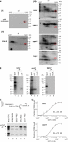
Figure 1.
Figure 1.
hnRNP A1 binds to the human apaf-1 and rhinovirus IRES. (Ai) Western immunoblot (αA1) analysis of hnRNP A1 after 2D gel electrophoresis of HeLa nuclear extract was done with the 4B10 mAb against hnRNP A1. (ii and iii) UV cross-linking of HeLa cell nuclear extracts (NE) with 32P-labeled RNAs corresponding to the 5′ UTRs of the FGF-2 mRNA (ii) or to the HRV, EMCV, apaf-1, and bag1 mRNAs (iii). UV-cross-linked proteins were analyzed by 2D gel electrophoresis. Molecular-weight markers in kilodaltons are indicated at the right-hand side of each gel. pI represents the isoelectric point. The red circle indicates the position of hnRNP A1 migration. (B) UV cross-linking of HeLa NE with 32P-labeled HRV, apaf-1 and EMCV 5′ UTR RNAs. The positions of protein molecular-weight markers in kilodaltons are indicated at the left-hand side of the gels. Immunoprecipitation (IP) of cross-linked RNA-protein complexes was performed with (αA1) or without (mock) the 4B10 mAb. (C) Streptavidin acrylamide bead precipitation of hnRNP A1 protein with biotinylated RNA. 35S-labeled in vitro-translated hnRNP A1 protein was incubated with biotinylated RNAs corresponding to the HRV and apaf-1 5′ UTRs. The biotinylated RNA–35S-protein complexes were then precipitated with streptavidin acrylamide beads and analyzed by SDS-PAGE. The RNAs used corresponded to the EMCV IRES (lane 3), the HRV IRES (lane 4), and the apaf-1 IRES (lane 5). Lane 1 corresponds to 15% of the input/assay; lane 2 corresponds to incubation of the proteins with the beads in the absence of RNA. Bands indicated with a star represent truncated forms of hnRNP A1 resulting from spurious translation initiation/termination events in RRL. Experiments were performed at least five times. (D) hnRNP A1 binding curve to the HRV and apaf-1 5′ UTR RNAs. Filter binding assays were carried out and evaluated as described in Materials and Methods. Filter-bound RNA is plotted as function of the protein concentration and corrected for the fraction of active protein. Curves were fitted to average data points of three independent experiments.
Figure 2.
Figure 2.
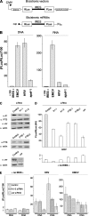
siRNA-mediated depletion of hnRNP A1 reduces HRV IRES activity. (A) Schematic representation of the bicistronic vectors or capped bicistronic mRNAs containing a 20-nucleotide-long poly (A) tail. CMV, cytomegalovirus promoter; RLuc, luciferase Renilla ORF; FLuc, luciferase firefly ORF. (B) The FLuc/RLuc activity ratio was measured in extracts of HeLa cells transfected with bicistronic vectors (DNA) (24 h posttransfection) or in vitro synthesized mRNAs (RNA) (6 h posttransfection). Bicistronic constructs containing the EMCV, the HRV, or the apaf-1 IRES were used. A no IRES construct was used as a control. The RLuc and FLuc values are shown in Supplemental Table S1. (C) The consequences of siRNA treatments were analyzed 48 h after siRNA transfection by Western immunoblotting with the indicated antibodies. α A1 corresponds to the monoclonal 4B10 antibody (Abcam), α PTB corresponds to a monoclonal Bb7 antibody provided by D. Black, α unr corresponds to a polyclonal antibody provided by H. Jacquemin-Sablon, and α actin corresponds to an actin polyclonal antibody (Sigma-Aldrich). The control lane corresponds to a transfection with a control siRNA. The RLuc and FLuc values are shown in Supplemental Table S2. (D) siRNA-mediated reductions in the expression of hnRNP A1, hnRNPI/PTB, or unr lead to various degrees of reduction in HRV IRES activity. The FLuc/RLuc ratio (AU, arbitrary units) driven by the HRV IRES or the no IRES construct were measured in extracts of HeLa cells transfected with bicistronic plasmids. The FLuc/RLuc ratio was arbitrarily set to 100 for extracts of HeLa cells transfected with the bicistronic plasmid containing the HRV IRES in the presence of the control siRNA. (E) siRNA-mediated reduction in the expression of hnRNP A1 reduces HRV IRES activity after DNA transfection, but not RNA transfection. The FLuc/RLuc ratio driven by the HRV IRES, the EMCV IRES or the no IRES construct was measured in extracts of HeLa cells transfected with bicistronic plasmids (DNA) or in vitro-synthesized mRNAs (RNA), and treated or not (control) with siRNAs targeting hnRNP A1 (A1 siRNA) or unr (unr siRNA). These experiments were performed at least three times.
Figure 3.
Figure 3.
Expression of a cytoplasmically restricted hnRNP A1 mutant leads to enhanced HRV IRES activity. (A) Schematic representation of the bicistronic reporter RNAs used to express a HA-tagged cytoplasmically restricted hnRNP A1 mutant (A1ΔM9) and a control protein (CAT), and to quantify rhinovirus IRES-mediated translation. The wild-type HRV IRES or the M5 mutant of the HRV IRES, which is reduced in its ability to interact with hnRNP A1 (see Supplemental Figure 1 and B), were inserted into the intercistronic space. (B) Streptavidin acrylamide precipitation of hnRNP A1 protein with biotinylated RNA. The RNAs used corresponded to a control RNA (lane 3), the wild-type HRV 5′ UTR (lane 4) or the M5 mutant of the HRV 5′ UTR (lane 5). Lane 1 corresponds to 15% of the input/assay; lane 2 corresponds to incubation of the proteins with the beads without RNA (C) Top, RPA using an antisense probe for the FLuc ORF (anti-FLuc). Bottom, Western-blot analysis of the expression of the mutant cytoplasmic hnRNP A1 by using an antibody against the HA tag (α HA). An actin Western blot was used as a loading control. (D) Immunocytochemistry experiments in HeLa cells were performed with the HA-tag antibody. (E) FLuc activity was measured in extracts of HeLa cells transfected with the various mRNAs, as indicated below the graph. This activity was normalized to the amount of transfected mRNAs measured by RPA experiments. The FLuc activity was arbitrarily set to 1 for the conditions corresponding to the transfection of the control CAT-HRV mRNA. These experiments were repeated three times for D, four times for C and E, and five times for B.
Figure 4.
Figure 4.
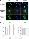
HRV IRES activity is stimulated upon rhinovirus infection. The human rhinovirus was added at a multiplicity of infection (MOI) of 0, 10, or 20 as indicated. After 1-h incubation, the virus inoculum was removed, and fresh medium was added to the culture. Cells were subsequently incubated for 10 h before harvesting and analysis. (A) hnRNP A1 accumulates in the cytoplasm in rhinovirus-infected HeLa cells. Immunostaining of noninfected or HRV-infected HeLa cells for hnRNP A1 (αA1) and the nuclear marker 4,6-diamidino-2-phenylindole (DAPI). The merged images (αA1/DAPI) show the presence of hnRNP A1 outside the nucleus in HRV-infected cells. (B) The FLuc/RLuc ratio was measured in extracts of HeLa cells transfected with in vitro synthesized bicistronic mRNAs containing either the HRV or the EMCV IRES. RNA transfections were performed 2 h before infection, whereas hnRNP A1 siRNA transfection was performed 48 h before infection. The RLuc and FLuc values are shown in Supplemental Table S3. This experiment was performed on three independent occasions (*p < 0.04).
Figure 5.
Figure 5.
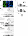
UVC-dependent cytoplasmic relocalization of hnRNP A1 limits apaf-1 mRNA translation. (A) hnRNP A1 accumulates in the cytoplasm in UVC-irradiated 293T cells. Immunostaining of control 293T cells (without UVC) or UVC irradiated (UVC: 300 J/m2) for hnRNP A1 (αA1) and the nuclear marker DAPI. The merged images (αA1/DAPI) show the presence of hnRNP A1 outside the nucleus in UVC-irradiated cells. (B) Representative polysome distribution profiles obtained after centrifugation of cytoplasmic lysates over sucrose gradients. Lysates were prepared from either irradiated cells (8 h after receiving 300 J/m2 UVC) (ii and iii) or nonirradiated cells (i). Irradiated cells were either transfected with siRNA targeting hnRNP A1 (iii) or left untreated (ii). From left to right, fractions contained ribosome subunits or single ribosomes (fractions 1–5) or polysomes of increasing molecular weights (fractions 6–13). From each fraction, RNA was prepared for agarose gel analysis of 18S and 28S rRNAs and for Northern blot of actin and apaf-1 mRNAs. This representative experiment was repeated three times. (C) Quantifications of the Northern blots from B. The percentages of the apaf-1 and actin mRNAs in each fraction are shown on two separated histograms. (D) The efficiency of siRNA treatment was analyzed by Western immunoblotting as in Figure 2C.
Figure 6.
Figure 6.
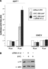
UVC-dependent cytoplasmic relocalization of hnRNP A1 limits apaf-1 IRES activity. (A) The FLuc and RLuc activities of transfected bicistronic mRNAs containing the apaf-1 and EMCV IRES were measured in extracts of 293T cells left untreated, irradiated with UVC, or irradiated with UVC after siRNA-mediated depletion of hnRNP A1. (B) The efficiency of siRNA treatment was analyzed by Western immunoblotting as in Figure 2C. This set of experiments was repeated at three different occasions.
Figure 7.
Figure 7.
Expression of a cytoplasmically restricted hnRNP A1 mutant limits the UVC-triggered activation of apaf-1 IRES activity. (A) Schematic representation of the bicistronic reporter RNAs used to express a HA-tagged cytoplasmically restricted hnRNP A1 mutant (A1ΔM9) or a control protein (CAT) and quantify apaf-1 IRES-mediated translation. (B) RPA using an antisense probe for the FLuc ORF (anti-FLuc). (C) The FLuc activities were measured in extracts of transfected 293T cells that were UVC-irradiated or nonirradiated, as indicated. The two mRNAs were used, as indicated. This activity was normalized to the amount of transfected mRNAs measured by RPA experiments. The FLuc activity was arbitrarily set to 1 for the condition corresponding to the transfection of the control CAT-apaf mRNA in non-UVC exposed cells. This experiment was repeated three times.

Similar articles

Cited by

References

    1. Allemand E., Guil S., Myers M., Moscat J., Caceres J. F., Krainer A. R. Regulation of heterogenous nuclear ribonucleoprotein A1 transport by phosphorylation in cells stressed by osmotic shock. Proc. Natl. Acad. Sci. USA. 2005;102:3605–3610. - PMC - PubMed
    1. Bedard K. M., Daijogo S., Semler B. L. A nucleo-cytoplasmic SR protein functions in viral IRES-mediated translation initiation. EMBO J. 2007;26:459–467. - PMC - PubMed
    1. Bonnal S., Boutonnet C., Prado-Lourenco L., Vagner S. IRESdb: the Internal Ribosome Entry Site database. Nucleic Acids Res. 2003;31:427–428. - PMC - PubMed
    1. Bonnal S., Pileur F., Orsini C., Parker F., Pujol F., Prats A. C., Vagner S. Heterogeneous nuclear ribonucleoprotein A1 is a novel internal ribosome entry site trans-acting factor that modulates alternative initiation of translation of the fibroblast growth factor 2 mRNA. J. Biol. Chem. 2005;280:4144–4153. - PubMed
    1. Boussadia O., Niepmann M., Creancier L., Prats A. C., Dautry F., Jacquemin-Sablon H. Unr is required in vivo for efficient initiation of translation from the internal ribosome entry sites of both rhinovirus and poliovirus. J. Virol. 2003;77:3353–3359. - PMC - PubMed

Publication types

MeSH terms

Substances

LinkOut - more resources