. 2007 Oct 2;104(40):15591-8.
doi: 10.1073/pnas.0704219104. Epub 2007 Sep 26.

What a difference a decade makes: insights into translesion DNA synthesis

Affiliations

What a difference a decade makes: insights into translesion DNA synthesis

Wei Yang et al. Proc Natl Acad Sci U S A. .

Abstract

Living organisms are continually under attack from a vast array of DNA-damaging agents that imperils their genomic integrity. As a consequence, cells possess an army of enzymes to repair their damaged chromosomes. However, DNA lesions often persist and pose a considerable threat to survival, because they can block the cell's replicase and its ability to complete genome duplication. It has been clear for many years that cells must possess a mechanism whereby the DNA lesion could be tolerated and physically bypassed. Yet it was only within the past decade that specialized DNA polymerases for "translesion DNA synthesis" or "TLS" were identified and characterized. Many of the TLS enzymes belong to the recently described "Y-family" of DNA polymerases. By possessing a spacious preformed active site, these enzymes can physically accommodate a variety of DNA lesions and facilitate their bypass. Flexible DNA-binding domains and a variable binding pocket for the replicating base pair further allow these TLS polymerases to select specific lesions to bypass and favor distinct non-Watson-Crick base pairs. Consequently, TLS polymerases tend to exhibit much lower fidelity than the cell's replicase when copying normal DNA, which results in a dramatic increase in mutagenesis. Occasionally this can be beneficial, but it often speeds the onset of cancer in humans. Cells use both transcriptional and posttranslational regulation to keep these low-fidelity polymerases under strict control and limit their access to a replication fork. Our perspective focuses on the mechanistic insights into TLS by the Y-family polymerases, how they are regulated, and their effects on genomic (in)stability that have been described in the past decade.


Conflict of interest statement

The authors declare no conflict of interest.

Figures

Fig. 1.
Fig. 1.
Structural domains of the Y-family polymerases. The polymerase domain is labeled in red (palm), blue (finger), green (thumb), purple (LF), and yellow (N-terminal addition in polκ and Rev1). The regulatory units are color- and shape-coded as indicated at the bottom of the figure. UBM stands for ubiquitin-binding motif, UBZ for ubiquitin-binding zinc finger, and BRCT for Brca1 C-terminal domain.
Fig. 2.
Fig. 2.
Structural comparison of T7 DNA polymerase (A-family) (PDB ID code 1T7P) and Dpo4 (Y-family) (PDB ID code 2AGQ). (A) The polymerase domain is shown in the same colors as in Fig. 1. Thioredoxin (wheat) enhances the processivity of T7 DNA pol. DNA is shown in yellow (primer) and brown (template) tubes, the two metal ions as cyan spheres, and the incoming nucleotide (only visible in Dpo4) as silver and multicolored sticks. (B) Diagrams of the conformational change of a helix (solid blue rectangle) in the finger domain of T7 DNA pol upon binding of a correct incoming nucleotide (dNTP). Movement of the helix is indicated by a gray arrow. The reactants and catalysts are snug in the closed active site. (C) Illustration of the flexible LF and thumb domains of Y-family polymerases, which facilitate the movement of the template–primer duplex. The spacious and open active site also allows multiple conformations of the dNTP (as diagramed in the upper right corner) and makes it difficult to align the 3′-OH of the primer strand, dNTP, metal ions, and catalytic carboxylates.
Fig. 3.
Fig. 3.
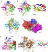
Structural and biochemical features of individual Y-family polymerases. (A) Dpo4 bypassing an abasic lesion (PDB ID code 1S0N). The polymerase domain is colored as in Fig. 1, and the red arrow points at the looped-out abasic site analog. The nucleotide 5′ to the abasic site serves as the template base to direct nucleotide incorporation. The dNTP is shown in sticks, and the two metal ions are shown as purple spheres. (B) Rev1 complexed with DNA (PDB ID code 2AQ4). The overall structures of Rev1 and Dpo4 are superimposable, including the incoming dNTP and metal ions. But the N-terminal region (shown in yellow) of Rev1 displaces the template base (highlighted in orange), and an Arg side chain is inserted in its place. A close-up view of the active site is shown on the right. The red arrow points at the Arg that forms two hydrogen bonds with dCTP. (C) Superposition of the structures of Dpo4 complexed with BPDE-dA-adducted DNA (PDB ID code 1S0M) (shown in silver with the BPDE-dA highlighted in red) and polκ complexed with a normal DNA (PDB ID code 2OH2) (shown in cyan with the N-terminal three-helix insertion highlighted in yellow). The two proteins (in which the α-helices are represented by cylinders), DNAs, and dNTPs in particular are superimposable. The N-terminal addition of polκ (yellow) can partially shield the BPDE adduct in the major groove, which otherwise is exposed to solvent as in the complex with Dpo4. (D) The backside of the polκ–DNA ternary complex structure. The polymerase domain is shown in a molecular surface representation and colored as in Fig. 1. The crevice separates the LF (purple) and finger domains (blue) and also extends to the palm domain. The normal template base is shown as red sticks. If it were a BPDE-dG adduct, the BPDE moiety in the minor groove could be accommodated in the large crevice during the nucleotide insertion step as well as the subsequent primer extension. (E) A close-up view of the polη active site (PDB ID code 1JIH). A CPD-containing DNA, dATP paired with the 3′ T of the CPD (shown as red sticks) and two metal ion (purple spheres) are borrowed from the Dpo4–CPD complex structure (PDB ID code 1RYR) after superimposing the palm and finger domains of the two proteins. The R73 in yeast polη (R61 in human polη), which is proposed to stabilize the incoming dATP, is shown as cyan and blue sticks (with the red arrow pointing at it). (F) Comparison of polι (PDB ID code 2FLL, colored in orange) and Dpo4 (PDB ID code 2AGQ, colored in silver). The overall structures of the two polymerase–substrate ternary complexes are quite similar. The replicating base pair in Dpo4 (shown in cyan) is superimposable with those in polκ and Rev1 (Fig. 4 B and C), but it differs from that in polι (magenta) because of potential clashes with the large aliphatic side chains (L62, V64, and L78, shown in yellow) present in the finger domain of polι. Interestingly, the triphosphate moieties of dNTP (orange and red) are more or less superimposable between Dpo4 and polι. A close-up view of the superimposed active sites is shown on the right.
Fig. 4.
Fig. 4.
A composite active site of the Y-family polymerases in stereoview. The 3′-end nucleotide of the primer strand (pale yellow), the template nucleotide (orange), the incoming dNTP [yellow (C)/blue (N)/red (O)], the three catalytic carboxylates [magenta (C)/red (O)], the nearby carbonyl group [F8(O)] that coordinates one metal ion, and the conserved residues interacting with dNTP [light blue (C)/blue (N)/red (O)] are shown as sticks, and the two metal ions are shown as green spheres. These conserved residues are labeled according to Dpo4 for convenience. K77 of polι that stabilizes the incoming dNTP and M135 of polκ that stabilizes the template base are shown in gray/blue (N)/brown (S) sticks. These residues are replaced by Ala's in Dpo4. The 3′-OH group (indicated as a red “o”) is usually absent in the crystal structures for the purpose of capturing enzyme–substrate complexes.
Fig. 5.
Fig. 5.
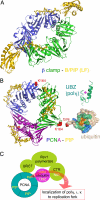
Access of Y-family polymerases to a replication fork is regulated by posttranslational modification and protein–protein interactions. (A) Ribbon diagram of E. coli polIV C-terminal region (including the LF domain and B/PIP) complexed with the β-clamp (PDB ID code 1UNN). The two subunits of β clamp are shown in green and blue, and polIV is shown in yellow. The B/PIP of polIV is represented by a stick model. (B) Interactions among PCNA–PIP (represented by p21, PDB ID code 1AXC), ubiquitin, UBZ, and UBM. The trimeric PCNA is shown in blue, green, and purple ribbon diagram. The PIP peptide from p21 is shown as yellow sticks. When ubiquitinated, PCNA is covalently linked through its K164 (represented by red spheres) with G76 (highlighted in red) of ubiquitin (PDB ID code 2G45). Ubiquitin is shown in molecular surface representation, the conserved I44 is highlighted in orange, and the surrounding areas that have been mapped to interact with UBZ and UBM are highlighted in green and blue, respectively. The NMR structure of the polη UBZ (PDB ID code 2I5O) is shown in green ribbon diagrams, and a magenta sphere represents the zinc ion. (C) A cartoon summarizing the protein–protein interactions of eukaryotic Y-family polymerases. Rev1 (pea green), polη, polι, and polκ each interact with PCNA (cyan) and ubiquitin (magenta), and the C-terminal region of Rev1 interacts with polη, polι, and polκ (collectively represented by the curvy red arrow). The multilayered interactions occur in response to DNA damage and may allow polη, polι, and polκ to be recruited to replication forks.

References

    1. Kato T, Shinoura Y. Mol Gen Genet. 1977;156:121–131. - PubMed
    1. Steinborn G. Mol Gen Genet. 1978;165:87–93. - PubMed
    1. Lemontt JF. Genetics. 1971;68:21–33. - PMC - PubMed
    1. Lawrence CW, Christensen R. Genetics. 1976;82:207–232. - PMC - PubMed
    1. Bridges BA, Woodgate R. Proc Natl Acad Sci USA. 1985;82:4193–4197. - PMC - PubMed

Publication types