

CXCR7 (RDC1) promotes breast and lung tumor growth in vivo and is expressed on tumor-associated vasculature
- PMID: 17898181
- PMCID: PMC1994579
- DOI: 10.1073/pnas.0610444104
CXCR7 (RDC1) promotes breast and lung tumor growth in vivo and is expressed on tumor-associated vasculature
Abstract
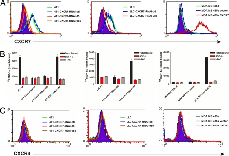
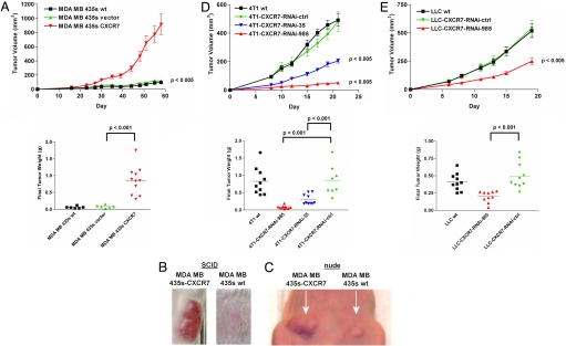
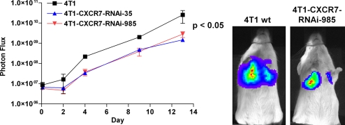
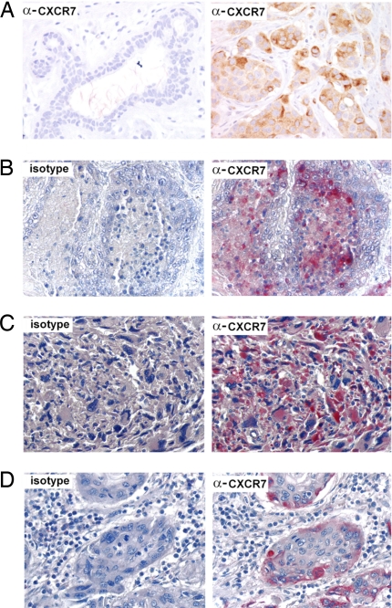
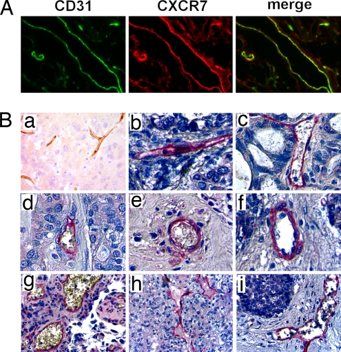
Chemokines and chemokine receptors have been posited to have important roles in several common malignancies, including breast and lung cancer. Here, we demonstrate that CXCR7 (RDC1, CCX-CKR2), recently deorphanized as a chemokine receptor that binds chemokines CXCL11 and CXCL12, can regulate these two common malignancies. Using a combination of overexpression and RNA interference, we establish that CXCR7 promotes growth of tumors formed from breast and lung cancer cells and enhances experimental lung metastases in immunodeficient as well as immunocompetent mouse models of cancer. These effects did not depend on expression of the related receptor CXCR4. Furthermore, immunohistochemistry of primary human tumor tissue demonstrates extensive CXCR7 expression in human breast and lung cancers, where it is highly expressed on a majority of tumor-associated blood vessels and malignant cells but not expressed on normal vasculature. In addition, a critical role for CXCR7 in vascular formation and angiogenesis during development is demonstrated by using morpholino-mediated knockdown of CXCR7 in zebrafish. Taken together, these data suggest that CXCR7 has key functions in promoting tumor development and progression.
Conflict of interest statement
The authors declare no conflict of interest.
Figures
Similar articles
-
Scavenging of CXCL12 by CXCR7 promotes tumor growth and metastasis of CXCR4-positive breast cancer cells.Oncogene. 2012 Nov 8;31(45):4750-8. doi: 10.1038/onc.2011.633. Epub 2012 Jan 23. Oncogene. 2012. PMID: 22266857 Free PMC article.
-
CXCL12-CXCR7 axis is important for tumor endothelial cell angiogenic property.Int J Cancer. 2015 Dec 15;137(12):2825-36. doi: 10.1002/ijc.29655. Epub 2015 Jul 2. Int J Cancer. 2015. PMID: 26100110
-
C-X-C motif chemokine 12/C-X-C chemokine receptor type 7 signaling regulates breast cancer growth and metastasis by modulating the tumor microenvironment.Breast Cancer Res. 2014 May 29;16(3):R54. doi: 10.1186/bcr3665. Breast Cancer Res. 2014. PMID: 24886617 Free PMC article.
-
Biological/pathological functions of the CXCL12/CXCR4/CXCR7 axes in the pathogenesis of bladder cancer.Int J Clin Oncol. 2017 Dec;22(6):991-1000. doi: 10.1007/s10147-017-1187-x. Epub 2017 Oct 11. Int J Clin Oncol. 2017. PMID: 29022185 Review.
-
CXCR7 impact on CXCL12 biology and disease.Trends Mol Med. 2013 Jan;19(1):12-22. doi: 10.1016/j.molmed.2012.10.004. Epub 2012 Nov 12. Trends Mol Med. 2013. PMID: 23153575 Review.
Cited by
-
CXCR7 protein expression correlates with elevated mmp-3 secretion in breast cancer cells.Oncol Lett. 2010 Sep;1(5):845-847. doi: 10.3892/ol_00000149. Epub 2010 Sep 1. Oncol Lett. 2010. PMID: 22966392 Free PMC article.
-
Opposing roles of CXCR4 and CXCR7 in breast cancer metastasis.Breast Cancer Res. 2011;13(6):R128. doi: 10.1186/bcr3074. Epub 2011 Dec 9. Breast Cancer Res. 2011. PMID: 22152016 Free PMC article.
-
Inhibition of Angiogenesis by MiR-524-5p through Suppression of AKT and ERK Activation by Targeting CXCR7 in Colon Cancer Cells.J Oncol. 2022 Nov 10;2022:7224840. doi: 10.1155/2022/7224840. eCollection 2022. J Oncol. 2022. PMID: 36405246 Free PMC article.
-
The Signaling Duo CXCL12 and CXCR4: Chemokine Fuel for Breast Cancer Tumorigenesis.Cancers (Basel). 2020 Oct 21;12(10):3071. doi: 10.3390/cancers12103071. Cancers (Basel). 2020. PMID: 33096815 Free PMC article. Review.
-
Cytoplasmic CXCR4 expression in breast cancer: induction by nitric oxide and correlation with lymph node metastasis and poor prognosis.BMC Cancer. 2008 Nov 23;8:340. doi: 10.1186/1471-2407-8-340. BMC Cancer. 2008. PMID: 19025611 Free PMC article.
References
-
- Balkwill F. Nat Rev Cancer. 2004;4:540–550. - PubMed
-
- Hartmann T, Burger M, Burger J. J Biol Regul Homeost Agents. 2004;18:126–130. - PubMed
-
- Luker K, Luker G. Cancer Lett. 2006:23830–41. - PubMed
-
- Allinen M, Beroukhim R, Cai L, Brennan C, Lahti-Domenici J, Huang H, Porter D, Hu M, Chin L, Richardson A, et al. Cancer Cell. 2004;6:17–32. - PubMed
-
- Orimo A, Gupta P, Sgroi D, Arenzana-Seisdedos F, Delaunay T, Naeem R, Carey V, Richardson A, Weinberg R. Cell. 2005;121:335–348. - PubMed
Publication types
MeSH terms
Substances
Grants and funding
LinkOut - more resources
Full Text Sources
Other Literature Sources
Medical
Molecular Biology Databases
