. 2007 Nov;14(11):1400-8.
doi: 10.1128/CVI.00299-07. Epub 2007 Sep 26.

Use of protein antigens for early serological diagnosis of leprosy

Affiliations

Use of protein antigens for early serological diagnosis of leprosy

Malcolm S Duthie et al. Clin Vaccine Immunol. 2007 Nov.

Abstract

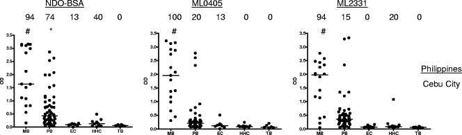
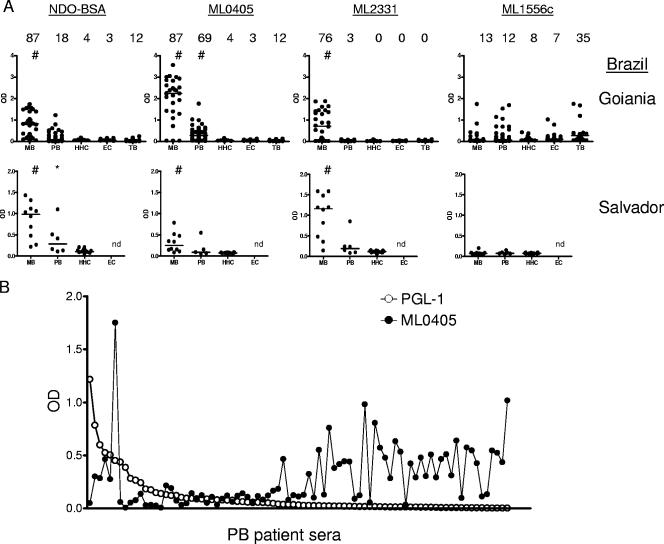
Leprosy is a chronic and debilitating human disease caused by infection with the Mycobacterium leprae bacillus. Despite the marked reduction in the number of registered worldwide leprosy cases as a result of the widespread use of multidrug therapy, the number of new cases detected each year remains relatively stable. This indicates that M. leprae is still being transmitted and that, without earlier diagnosis, M. leprae infection will continue to pose a health problem. Current diagnostic techniques, based on the appearance of clinical symptoms or of immunoglobulin M (IgM) antibodies that recognize the bacterial phenolic glycolipid I, are unable to reliably identify early-stage leprosy. In this study we examine the ability of IgG within leprosy patient sera to bind several M. leprae protein antigens. As expected, multibacillary leprosy patients provided stronger responses than paucibacillary leprosy patients. We demonstrate that the geographic locations of the patients can influence the antigens they recognize but that ML0405 and ML2331 are recognized by sera from diverse regions (the Philippines, coastal and central Brazil, and Japan). A fusion construct of these two proteins (designated leprosy IDRI diagnostic 1 [LID-1]) retained the diagnostic activity of the component antigens. Upon testing against a panel of prospective sera from individuals who developed leprosy, we determined that LID-1 was capable of diagnosing leprosy 6 to 8 months before the onset of clinical symptoms. A serological diagnostic test capable of identifying and allowing treatment of early-stage leprosy could reduce transmission, prevent functional disabilities and stigmatizing deformities, and facilitate leprosy eradication.


Figures

FIG. 1.
FIG. 1.
Sera from Filipino leprosy patients react with recombinant M. leprae antigens. Sera from clinically diagnosed MB and PB leprosy patients, EC individuals, and HHC of MB leprosy patients were assessed against NDO-BSA, ML0405, and ML2331. NDO-BSA reactivity was assessed by IgM binding, and protein reactivity was assessed by IgG binding. Sera were from Cebu City, Philippines. Each point represents an individual serum sample, and the median is represented by the line. The number above each data set is the percent positive responses. *, P < 0.05; #, P < 0.001 versus EC.
FIG. 2.
FIG. 2.
ML0405 constructs react with MB leprosy patient sera. Different ML0405 constructs were created and expressed as recombinant proteins. The schematic diagram shows the sequence alignment of each of these constructs, with the deleted regions indicated by the line. Each construct was tested for IgG reactivity by ELISA with individual Filipino MB leprosy patient sera (n = 18) or EC sera (n = 6). *, P < 0.05; #, P < 0.001 versus EC.
FIG. 3.
FIG. 3.
M. leprae proteins react with PB leprosy patient sera. (A to C) Antibody reactivities of sera from a pool of clinically diagnosed MB leprosy patients, from a pool of negative control individuals, and from 46 clinically diagnosed PB leprosy patients were assessed against NDO-BSA and ML0405 (A), ML2331 (B), and ML1556c (C). NDO-BSA reactivity was assessed by IgM binding and, for reference, is shown in each plot. Recombinant protein reactivity was assessed by IgG binding. The first open circle represents the value obtained for pooled MB sera, while the next open circle represents the reactivity of pooled EC sera; individual PB sera are then arranged along the x axis according to their responsiveness versus NDO-BSA. The dashed line indicates the point at which diagnosis by NDO-BSA reactivity becomes unclear. ML1556c reacts with PB leprosy patient sera. (D) IgG reactivities of ML1556c with a small panel of individual sera from EC, leprosy patients (MB and PB), and TB patients were determined by ELISA using samples from Cebu City, Philippines. Each point represents an individual serum sample, and the median is represented by the line.
FIG. 4.
FIG. 4.
Sera from Brazilian leprosy patients react with recombinant M. leprae antigens. Sera from clinically diagnosed MB and PB leprosy patients, EC individuals, and HHC of MB leprosy patients were assessed against NDO-BSA, ML0405, ML2331, and ML1556c. NDO-BSA reactivity was assessed by IgM binding, and protein reactivity was assessed by IgG binding. Sera were from Goiânia and Salvador (see Table 1). (A) Each point represents an individual serum sample, and the median is represented by the line. The number above each data set is the percent positive responses. *, P < 0.05; #, P < 0.001 versus EC. (B) To demonstrate complementarity, the individual PB sera from Goiânia are arranged along the x axis according to their responsiveness versus NDO-BSA and overlaid with the response of each serum to ML0405.
FIG. 5.
FIG. 5.
LID-1 retains reactivity with leprosy patient sera. (A) LID-1 (a fusion construct of ML0405 and ML2331), ML0405FL, ML0405Tr, and ML2331 reactivities were assessed by IgG binding in an ELISA with eight MB leprosy patient serum samples from Salvador and eight NEC serum samples. (B) Sera from clinically diagnosed Japanese MB and PB leprosy patients, and Japanese EC individuals, were assessed for IgG reactivities with LID-1, ML0405, and ML2331. Each point represents an individual serum sample, and the median is represented by the line. The number above each data set is the percent positive responses. *, P < 0.05; #, P < 0.001 versus EC.
FIG. 6.
FIG. 6.
LID-1 reactivity can diagnose leprosy before clinical symptoms. (A) LID-1 and NDO-BSA reactivities within sera from a prospective study conducted in Cebu City, Philippines, were assessed by either IgG or IgM binding in an ELISA. Sera were collected at a variety of times prior to the clinical diagnosis of MB leprosy in 11 patients and at a variety of times after the commencement of treatment. (B) Representative plots for individual patients are shown. (C) Sera were collected from 57 household contacts that did not develop clinical leprosy and were compared with single serum samples from each individual contact that developed leprosy (serum samples were collected within 3 months of clinical diagnosis). #, P < 0.001. (D) LID-1 and NDO-BSA reactivities within sera from a prospective study using 10 U.S.-based individuals who were immunized with BCG were assessed. Sera were collected at regular intervals following BCG immunization. The solid circle at day zero designates the reactivity of a leprosy patient serum sample that was included as a positive control.

References

    1. Anderson, H., B. Stryjewska, B. L. Boyanton, and M. R. Schwartz. 2007. Hansen disease in the United States in the 21st century: a review of the literature. Arch. Pathol. Lab. Med. 131:982-986. - PubMed
    1. Bakker, M. I., M. Hatta, A. Kwenang, P. Van Mosseveld, W. R. Faber, P. R. Klatser, and L. Oskam. 2006. Risk factors for developing leprosy—a population-based cohort study in Indonesia. Lepr. Rev. 77:48-61. - PubMed
    1. Buhrer, S. S., H. L. Smits, G. C. Gussenhoven, C. W. van Ingen, and P. R. Klatser. 1998. A simple dipstick assay for the detection of antibodies to phenolic glycolipid-I of Mycobacterium leprae. Am. J. Trop. Med. Hyg. 58:133-136. - PubMed
    1. Buhrer-Sekula, S., H. L. Smits, G. C. Gussenhoven, J. van Leeuwen, S. Amador, T. Fujiwara, P. R. Klatser, and L. Oskam. 2003. Simple and fast lateral flow test for classification of leprosy patients and identification of contacts with high risk of developing leprosy. J. Clin. Microbiol. 41:1991-1995. - PMC - PubMed
    1. Cartel, J. L., S. Chanteau, J. P. Boutin, R. Plichart, P. Richez, J. F. Roux, and J. H. Grosset. 1990. Assessment of anti-phenolic glycolipid-I IgM levels using an ELISA for detection of M. leprae infection in populations of the South Pacific Islands. Int. J. Lepr. Other Mycobact. Dis. 58:512-517. - PubMed

Publication types

MeSH terms